When it comes to studying large-sized organs or tissues, such as brains, kidneys, or even entire embryos, researchers often face a significant challenge in obtaining thin and consistent sections for analysis. Traditional sectioning methods may struggle to handle the size and delicate nature of these specimens. However, we at Precisionary Instruments have developed a powerful tool to overcome these challenges and achieve precise, high-quality sections from large-sized specimens. In this article, we will explore how vibratomes have revolutionized the process of sectioning large-sized organs and tissues, opening up new possibilities for research and discovery. Check out a test section of two hard boiled eggs:

In the world of tissue sectioning, there are instances when traditional methods fall short in accommodating the size and complexity of large organs, such as the brain. This is where large diameter tissue sectioning steps in, offering a transformative approach to studying these intricate structures. By employing advanced techniques and tools like vibratomes, researchers can now delve into the vast landscape of whole brain sectioning, enabling comprehensive analysis and uncovering valuable insights into neural circuitry, connectivity, and disease mechanisms.
Large diameter tissue sectioning has opened up exciting avenues in biomedical research, enabling scientists to delve deeper into the intricate structures of organs and tissues. This innovative technique allows for the study of whole brain anatomy and histology, as well as the generation of precision-cut slices of entire lung lobes or liver lobes. In this article, we will explore the experimental applications of large diameter tissue sectioning and how it is advancing our understanding of complex biological systems.
Large diameter tissue sectioning provides a unique opportunity to study the anatomy and histology of the entire brain. By sectioning the brain as a whole, researchers can gain comprehensive insights into its structure, organization, and connectivity. This approach allows for the mapping of neuronal circuits, the identification of cell types, and the characterization of brain regions, leading to a deeper understanding of brain function and neurological disorders.
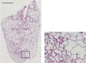
Large diameter tissue sectioning techniques have enabled the generation of precision-cut lung slices from entire lung lobes. These slices closely mimic the in vivo environment, allowing researchers to study lung physiology, airway dynamics, and tissue responses in a controlled ex vivo setting. These slices serve as valuable models for investigating lung diseases, drug screening, and assessing the efficacy of therapeutic interventions.
Similar to lung slices, large diameter tissue sectioning facilitates the generation of precision-cut slices from entire liver lobes. These slices provide a platform for studying liver anatomy, function, and disease. Researchers can examine hepatic cell interactions, investigate metabolic processes, and evaluate drug metabolism and toxicity. Precision-cut liver slices are also valuable for assessing liver regeneration, transplantation, and developing targeted therapies for liver diseases.
Large diameter tissue sectioning enables the study of organ-level interactions within complex biological systems. By sectioning multiple organs or tissues together, researchers can investigate the dynamic relationships and communication between different components of an organ system. This approach provides valuable insights into physiological processes, disease progression, and therapeutic interventions.
Large diameter tissue sectioning bridges the gap between basic research and clinical applications. The generation of large tissue sections allows for comprehensive analysis, including molecular profiling, immunohistochemistry, and imaging techniques. By correlating experimental findings with clinical data, researchers can better understand disease mechanisms, identify diagnostic biomarkers, and develop personalized treatment strategies.

The Compresstome VF-800-0Z vibratome has revolutionized the field of large diameter tissue sectioning, offering researchers a powerful and precise tool to obtain high-quality sections from complex organ systems. Developed with a focus on versatility and efficiency, this innovative vibratome enables scientists to explore the intricate details of large tissues with exceptional precision. In this article, we will delve into the features and benefits of the Compresstome VF-800-0Z, and explore how it has transformed the cutting of large diameter tissue sections.
The Compresstome VF-800-0Z is specifically designed to provide enhanced stability and control during the sectioning process. With its robust construction and advanced vibration technology, it minimizes tissue movement and vibrations, resulting in clean and precise cuts. This stability is crucial for maintaining the integrity of large tissue specimens and ensuring accurate sectioning.
One of the key advantages of the Compresstome VF-800-0Z is its ability to accommodate large tissue sizes. With an expanded cutting platform and adjustable specimen stages, this vibratome can handle a wide range of tissue dimensions. Whether it is whole organs, brain regions, or tissue constructs, researchers can confidently section large diameter tissues with ease, without compromising the quality of the sections.
Consistency is paramount when it comes to large diameter tissue sectioning, and the Compresstome VF-800-0Z delivers on this front. Equipped with advanced cutting features and customizable settings, researchers can achieve uniform section thickness throughout the entire specimen. This uniformity ensures reliable and reproducible results, enabling accurate analysis and interpretation of the tissue samples.
The Compresstome VF-800-0Z prioritizes user-friendliness, simplifying the sectioning process for researchers. Its intuitive interface and user-friendly controls allow for easy adjustment of cutting parameters, such as speed and amplitude, to suit specific tissue types and experimental requirements. This user-centric design streamlines the workflow and minimizes the learning curve, enabling researchers to achieve optimal results in less time.
The versatility of the Compresstome VF-800-0Z makes it an ideal choice for a wide range of applications. From neuroscience research to regenerative medicine and beyond, researchers can explore the intricacies of large diameter tissue sections with this adaptable vibratome. Its compatibility with various staining and imaging techniques further expands the possibilities for in-depth analysis and characterization of the tissues.
The Compresstome VF-800-0Z vibratome has emerged as a game-changer in the field of large diameter tissue sectioning. With its stability, versatility, and precision, researchers can confidently tackle the challenges posed by complex tissues, achieving high-quality sections for a myriad of applications. As the demand for large diameter tissue sectioning grows, the Compresstome VF-800-0Z continues to empower scientists, driving new discoveries and advancements in biomedical research. Check out a video of large diameter tissue cutting here with our Compresstome VF-800-0Z vibratome:
At Precisionary Instruments, we’re thrilled to shine a spotlight on innovative labs around the world that are advancing science in unique and meaningful ways. This
Artificial Cerebrospinal Fluid (ACSF) is an essential buffer solution widely used in electrophysiology experiments, particularly to keep acute brain slices alive during research. This solution closely mimics the natural cerebrospinal fluid found in the brain and is designed to maintain neuronal viability and function during experiments like patch-clamp recordings. In this article, we’ll explore the importance of ACSF, its composition, and practical tips to optimize its use.
You’ll hear back from us in one business day
© 2023 Copyright
*Academic discounts are only valid for customers in North America.
© 2023 copyright