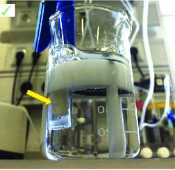
Oxygenation of a buffer solution for electrophysiology is important to maintain the viability of cells and tissues, as well as to prevent the formation of artifacts during recordings. Here are the general steps to oxygenate a buffer solution for electrophysiology:
Note that the specific protocol for oxygenation may vary depending on the type of buffer and the requirements of the experiment. Additionally, it is important to maintain sterility during the oxygenation process to avoid contamination of the buffer solution.

Yes, aerosolized buffer particles can potentially damage electrophysiology equipment, especially sensitive equipment such as microelectrodes, patch pipettes, and amplifiers. When buffer particles become aerosolized, they can settle on the surfaces of the equipment and cause damage over time. Additionally, buffer particles can also cause electrical noise and interfere with the accuracy of recordings.
To minimize the risk of equipment damage from aerosolized buffer particles, it is important to take the following precautions:
By taking these precautions, you can help protect your electrophysiology equipment from damage caused by aerosolized buffer particles and ensure accurate and reliable recordings.
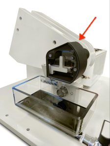
A Compresstome is a type of vibrating microtome that is commonly used for slicing thin sections of tissue for electrophysiology experiments. To maintain tissue viability during slicing, it is important to oxygenate the buffer solution used to immerse the tissue.
Some Compresstome models come with an oxygenation attachment that allows you to bubble oxygen into the buffer solution during the slicing process. This attachment typically consists of a tube that connects to a source of pure oxygen and a gas dispersion stone that is placed in the buffer solution.

The Compresstome VF-510-0Z is a type of vibrating microtome that is used for slicing thin sections of tissue for electrophysiology and other applications. The protective cover for the VF-510-0Z is an accessory that is designed to protect the user and the surrounding environment from the tissue and buffer solution that may be generated during the slicing process.
The protective cover is typically made of a clear acrylic material that allows the user to observe the slicing process while also providing a barrier to protect against splashes and other forms of contamination. The cover is easy to install and can be removed quickly for cleaning and maintenance.

At Precisionary Instruments, we’re thrilled to shine a spotlight on innovative labs around the world that are advancing science in unique and meaningful ways. This
Artificial Cerebrospinal Fluid (ACSF) is an essential buffer solution widely used in electrophysiology experiments, particularly to keep acute brain slices alive during research. This solution closely mimics the natural cerebrospinal fluid found in the brain and is designed to maintain neuronal viability and function during experiments like patch-clamp recordings. In this article, we’ll explore the importance of ACSF, its composition, and practical tips to optimize its use.
You’ll hear back from us in one business day
© 2023 Copyright
*Academic discounts are only valid for customers in North America.
© 2023 copyright