Mice are widely used as animal models in biomedical research due to their genetic, physiological, and behavioral similarities to humans. Mice are used to study a wide range of diseases and disorders, including cancer, diabetes, neurodegenerative diseases, and infectious diseases. They are also used for preclinical testing of drugs and other therapies.
In particular, the Compresstome® helps to section acute brain slices that are healthier, with a greater number of live neurons for electrophysiology, optogenetics, and calcium-imaging experiments. Brain slices cut with the Compresstome® are viable for up to twice as long as those made by other tissue slicers.
For fixed tissue experiments such as immunohistochemistry (IHC) or in-situ hybridization (ISH), the Compresstome® can section some slices as thin as 4um. Fresh slices may be cut down to as thin as 40um in thickness for organotypic culture or tumor slice studies.
Not sure which model is right for your needs?
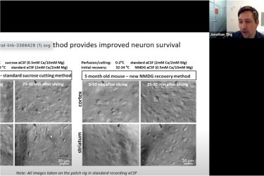

Jonathan T. Ting is an Assistant Investigator at the Allen Institute, where he joined in 2013 to provide electrophysiology expertise for the Human Cell Types program, and to develop functional assays on human ex vivo brain slides. In this webinar, Dr. Ting discusses which key steps in the brain slice process is most important and why, and challenges our conventional beliefs of slicing solutions and methodologies.
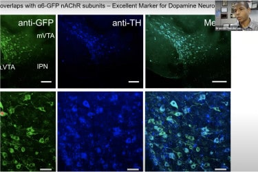

Assistant Professor in the Department of Biomedical Sciences at Marshall University’s Joan C. Edwards School of Medicine. In addition, Dr. Henderson and his focus on the role tobacco and vaping flavors play in addiction-related behaviors, and uses the Compresstome® vibrating microtome to make all of their acute brain slices for patch-clamp electrophysiology.
Dr Astero Klampatsa (PhD) is a Team Leader in Cancer Immunotherapy at the Institute of Cancer Research, London, UK and a Senior Lecturer in King’s College London, UK. She focuses on developing novel CAR T cell therapies for mesothelioma and lung cancer, as well as the immunobiology of these malignancies for identification of markers of response to immunotherapy. In this webinar, Dr. Klampatsa will discuss how the Compresstome® was used to create precision-cut tumor slices (PCTS) as an ex vivo model for immunotherapy research.

Often heralded as leaders in the field, the Allen Brain institute performs pioneering research on all manner of brain tissue. Working with brain tissue can often be as frustrating as it is rewarding. Slicing brain tissue presents many challenges. The tissue is a combination of soft and fibrous regions. For over a decade, researchers at the Allen Institute for Brain Science have been using the Compresstome® vibrating microtome to help give them better brain slices with increased longevity and reduced damage to surface neurons. This enables neuroscientists to have healthy neurons for patch-clamp electrophysiology experiments.

Researchers have used the Compresstome® in their procedure to section mouse embryo hypothalamus that has been injected with DNA and electroporated. This procedure demonstrates how it is possible to transfect nuclei in the hypothalamus region which are less accessible than those in superficial regions. Following this procedure additional experiments can be performed such as immunohistochemistry and in situ hybridization.


Explore how scientists use the Compresstome® vibrating microtome to create tissue slices that combine lipophilic dye tracing, whole mount in situ hybridization, immunohistochemistry, and histology to extract the maximal possible amount of data.


Dr. Wong shares how he built a custom-made Compresstome® for high-speed histological 3D imaging of whole organs like brains.


Dr. Chioccioli:


Dr. Fordahl will highlight how proinflammatory cytokines alter dopamine terminal function, and how increasing dietary fat intake may enhance microglial activity.


Dr. Greta Vargova is going to present their most recent publication, in which they show the link between locus coeruleus activity and behavioral flexibility.


Dr. Ransdell explores how the Compresstome vibrating microtome is used to produce healthy brain slices for electrophysiology. He studies adult Purkinje neurons in mouse cerebellar brain slices.


In this webinar, Dr. Wang will:
Godino A, Salery M, Durand-de Cuttoli R, Estill MS, Holt LM, Futamura R, Browne CJ, Mews P, Hamilton PJ, Neve RL, Shen L, Russo SJ, Nestler EJ. Transcriptional control of nucleus accumbens neuronal excitability by retinoid X receptor alpha tunes sensitivity to drug rewards. Neuron. 2023 May 3;111(9):1453-1467.e7. Epub 2023 Mar 7. PMID: 36889314; PMCID: PMC10164098. Download PDF
Li L, Durand-de Cuttoli R, Aubry AV, Burnett CJ, Cathomas F, Parise LF, Chan KL, Morel C, Yuan C, Shimo Y, Lin HY, Wang J, Russo SJ. Social trauma engages lateral septum circuitry to occlude social reward. Nature. 2023 Jan;613(7945):696-703. Epub 2022 Nov 30. PMID: 36450985; PMCID: PMC9876792. Download PDF
Wang C, Hyams B, Allen NC, Cautivo K, Monahan K, Zhou M, Dahlgren MW, Lizama CO, Matthay M, Wolters P, Molofsky AB, Peng T. Dysregulated lung stroma drives emphysema exacerbation by potentiating resident lymphocytes to suppress an epithelial stem cell reservoir. Immunity. 2023 Mar 14;56(3):576-591.e10. Epub 2023 Feb 22. PMID: 36822205. Download PDF
You’ll hear back from us in one business day
© 2023 Copyright
*Academic discounts are only valid for customers in North America.
© 2023 copyright