Visual guides to help you succeed with the Compresstome® vibrating microtomes, rotary microtomes, and all aspects of tissue sectioning
Our short training videos here detail everything you need to get started using your Compresstome® vibrating microtome. Explore how to unpack, set up, and begin preparing tissue samples with your new vibratome.
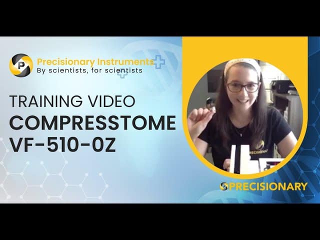
Step-by-step training video
Step-by-step training video
These short videos showcase the key features of each of our rotary microtome models. See which microtome best suites your research and clinical needs, and watch them operate in action!

Fully automated model
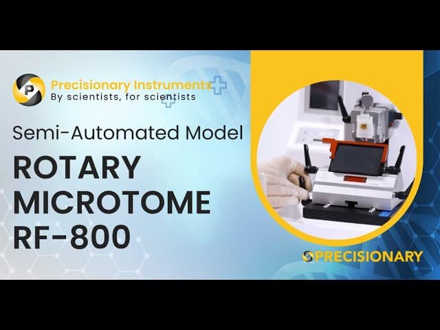
Semi-automated model
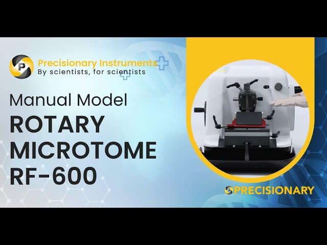
Manual model
View our in-depth training videos for each rotary microtome. We teach you to understand the parts of your rotary microtome, how to operate all microtome components, and show you how to set up tissue samples for cutting.
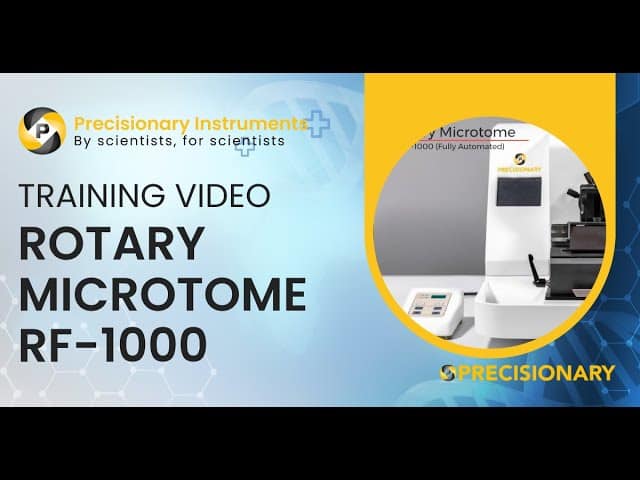
Fully automated model
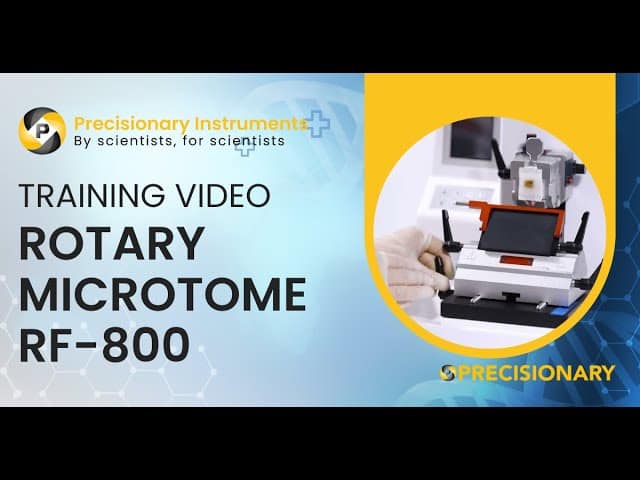
Semi-automated model
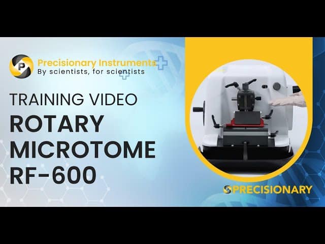
Manual model
Our Compresstome® vibrating microtomes and rotary microtomes have been used worldwide for almost 20 years. Hundreds of publications have been made possible due to our vibratomes and microtomes. Explore these research cases where experts use our products to make tissue slices for their experiments.

Often heralded as leaders in the field, the Allen Brain institute performs pioneering research on all manner of brain tissue. Working with brain tissue can often be as frustrating as it is rewarding. Slicing brain tissue presents many challenges. The tissue is a combination of soft and fibrous regions. For over a decade, researchers at the Allen Institute for Brain Science have been using the Compresstome® vibrating microtome to help give them better brain slices with increased longevity and reduced damage to surface neurons. This enables neuroscientists to have healthy neurons for patch-clamp electrophysiology experiments.

Researchers have used the Compresstome® in their procedure to section mouse embryo hypothalamus that has been injected with DNA and electroporated. This procedure demonstrates how it is possible to transfect nuclei in the hypothalamus region which are less accessible than those in superficial regions. Following this procedure additional experiments can be performed such as immunohistochemistry and in situ hybridization.


Explore how scientists use the Compresstome® vibrating microtome to create tissue slices that combine lipophilic dye tracing, whole mount in situ hybridization, immunohistochemistry, and histology to extract the maximal possible amount of data.


Fresh tissue can vary wildly in its level of difficulty to cut, due to a variety of factors like tissue type, and maturity of the animal (myelination). Often with other vibrating microtomes, they struggle to handle highly myelinated tissue or very soft neonatal tissue. The compression effect, along with multiple points of adjustment (speed, oscillation, and agarose concentration) enables our instrument to better handle “difficult” to cut tissue. The Compresstome® isn’t just able to cut thinner than the competition, we believe that the evidence shows that we also provide higher quality cuts that preserve cell surface structures and help increase the number of healthy to dead cells. Researchers at University of Minnesota use a Compresstome® to section live tissue in their procedure to locate, quantify, and phenotype antigen-specific CD8 T cells.


The footage above shows how neuroscientists use the Compresstome® tissue slicer to obtain brain slices for imaging studies. When using the Compresstome® tissue is embedded inside one of the specimen tubes. The specimen tube is made of two pieces: the inner plastic plunger and the outer metal tube. The inner plastic plunger is where you will mount your sample. The plunger is drawn down for your tissue sample to be embedded inside the metal tube. Please visit our website to learn more about our unique tissue slicers.


The Compresstome® has been widely used by researchers worldwide for making precision-cut lung slices (PCLS). The Compresstome® uses agarose embedding prior to slicing to allow for the preservation of open alveoli and better tissue compliance. The video above shows Compresstome® sectioning PCLS for immunostaining to visualize the localization of various immune cell types in the lung. This protocol can be extended to visualize the location and function of many different cell types under a variety of conditions.
You’ll hear back from us in one business day
© 2023 Copyright
*Academic discounts are only valid for customers in North America.
© 2023 copyright