

Key Takeaways
- Elevated CO2 (hypercapnia), common in advanced COPD, actively drives airway smooth muscle constriction rather than simply being a downstream symptom of disease severity.
- Hypercapnia activates ERK, which triggers calcium release and boosts STIM1 expression, amplifying calcium signaling that drives airway smooth muscle contraction.
- Precision-cut lung slices, prepared on a Compresstome tissue slicer, confirmed that STIM1 is required for hypercapnia-induced airway constriction in intact living tissue, not just isolated cells.
- Genetic variants that reduce STIM1 expression are enriched in COPD patients and normally protect against airway resistance, but that protection is lost under hypercapnia, pointing to a gene-environment interaction relevant to disease progression.
For patients with advanced chronic obstructive pulmonary disease (COPD), elevated blood carbon dioxide levels — a condition known as hypercapnia — have long been associated with worse clinical outcomes. But is hypercapnia merely a passive marker of how severe the disease has become, or is it actively making things worse? A landmark new study published in JCI Insight by Shigemura et al. (2026) answers that question decisively: hypercapnia is an active environmental stressor that directly drives airway smooth muscle constriction through a precisely defined molecular signaling cascade — the CO₂–ERK–STIM1–SOCE axis. This work, led by a large multi-institutional team including researchers at Northwestern University, Justus Liebig University Giessen, and University College Dublin, reshapes our understanding of hypercapnic COPD and opens the door to targeted therapeutic strategies.
The Unresolved Question: Does Elevated CO₂ Itself Tighten the Airways?
Clinicians have known for decades that COPD patients with hypercapnia fare worse than those who maintain normal CO₂ levels. Yet the prevailing view treated hypercapnia as a downstream consequence of severe airflow limitation and respiratory muscle fatigue — essentially a symptom, not a cause. There were hints in the literature that elevated CO₂ could affect smooth muscle biology, but the intracellular mechanisms connecting a rise in CO₂ to sustained airway smooth muscle (ASM) contraction remained poorly defined. In particular, it was unclear how hypercapnia engaged calcium signaling — the master regulator of smooth muscle contractility — and whether these effects were clinically meaningful at the CO₂ levels actually seen in patients, not just in extreme experimental conditions.
This knowledge gap mattered enormously. If hypercapnia is merely a bystander, then the clinical priority is to treat the underlying lung disease and accept CO₂ elevation as an inevitable marker of severity. But if elevated CO₂ is actively constricting airways, then reducing CO₂ itself — for instance through non-invasive ventilation — becomes a direct therapeutic intervention, and the molecular pathway it engages becomes a drug target.
A Triphasic Calcium Response and the ERK–STIM1 Signaling Cascade
Using a sophisticated combination of single-cell live calcium imaging (GCaMP6 and Fura-2-AM reporters), molecular biology, and genetic approaches, Shigemura and colleagues mapped the complete signaling pathway from CO₂ exposure to sustained ASM contraction. Their findings reveal a remarkably orchestrated triphasic calcium response in airway smooth muscle cells exposed to hypercapnia:
- Phase 1: An initial dip in intracellular calcium associated with CO₂-driven acidification.
- Phase 2: A surge of calcium released from the sarcoplasmic reticulum (SR), driven by ERK-dependent phosphorylation of the inositol 1,4,5-trisphosphate receptor (InsP3R).
- Phase 3: Sustained store-operated calcium entry (SOCE) through STIM1/Orai1 channels at SR–plasma membrane junctions, maintained by both rapid post-translational modification and longer-term transcriptional upregulation of STIM1.
The mechanistic elegance is striking. ERK activation serves as the critical hub: it simultaneously phosphorylates InsP3R to deplete SR calcium stores (triggering Phase 2), phosphorylates STIM1 at serine residues 575, 608, and 621 to promote its translocation into puncta that couple to Orai1 channels (initiating Phase 3), and drives nuclear translocation of the transcription factor c-Fos, which then increases STIM1 gene transcription — ensuring that the SOCE response is not only initiated but amplified and sustained over time.
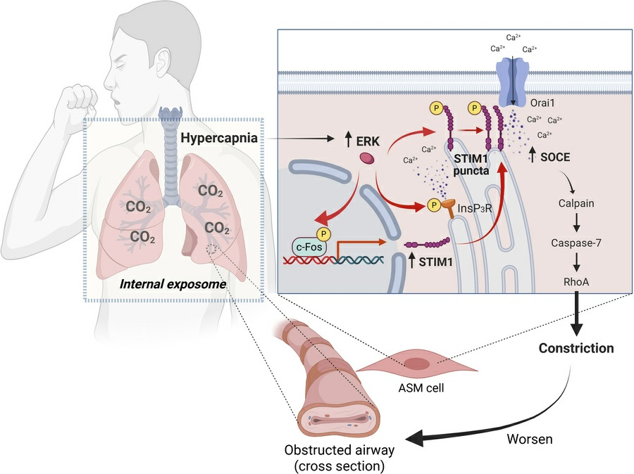
Figure 1. How high CO2 levels trigger a molecular cascade in airway muscle cells—boosting calcium signaling and causing airway constriction.
Crucially, these responses were not limited to supraphysiological CO₂ levels (~120 mmHg) used in many prior experimental setups. The team demonstrated that the same signaling axis operates at clinically relevant hypercapnia (50–60 mmHg) — the levels actually encountered in COPD patients — lending immediate translational weight to the findings.
From Molecular Signaling to Airway Constriction in Precision-Cut Lung Slices
Demonstrating a signaling cascade in cultured cells is essential, but the critical test of any proposed contractile mechanism is whether it drives actual airway narrowing in intact tissue. This is where precision tissue sectioning became indispensable to the study.
The researchers generated 100 µm precision-cut lung slices (PCLS) from mouse lungs using the Precisionary Instruments Compresstome® VF-300 vibrating microtome. At this thickness, small airways with intact smooth muscle layers are preserved within the slice, and individual airways can be monitored in real time as they constrict or relax in response to pharmacologic or environmental stimuli. The Compresstome’s agarose-compression approach was particularly important here: mouse lung tissue is exceptionally delicate, with thin-walled airways surrounded by compliant parenchyma that can easily tear or compress during sectioning. Traditional vibratome cutting can introduce mechanical artifact that disrupts the smooth muscle layer or distorts airway geometry — either of which would compromise a functional contraction assay. The Compresstome’s stabilization of the tissue within the agarose matrix during sectioning minimizes these artifacts, preserving the structural and functional integrity needed for live airway contraction measurements.
Using this PCLS platform, the team showed that hypercapnia significantly enhanced airway constriction in wild-type mouse lung slices. The functional requirement for STIM1 was then confirmed through two complementary loss-of-function strategies: shRNA knockdown of STIM1 in cultured ASM cells reduced hypercapnia-induced contractile signaling markers (calpain activity, caspase-7 cleavage, and RhoA activation), and smooth muscle–specific STIM1 knockout mice (SMMHC-CreERT2; Stim1fl/fl, tamoxifen-induced) showed markedly attenuated airway constriction in PCLS under hypercapnic conditions. Pharmacologic inhibition with BTP-2 and 2-APB further corroborated the genetic findings.
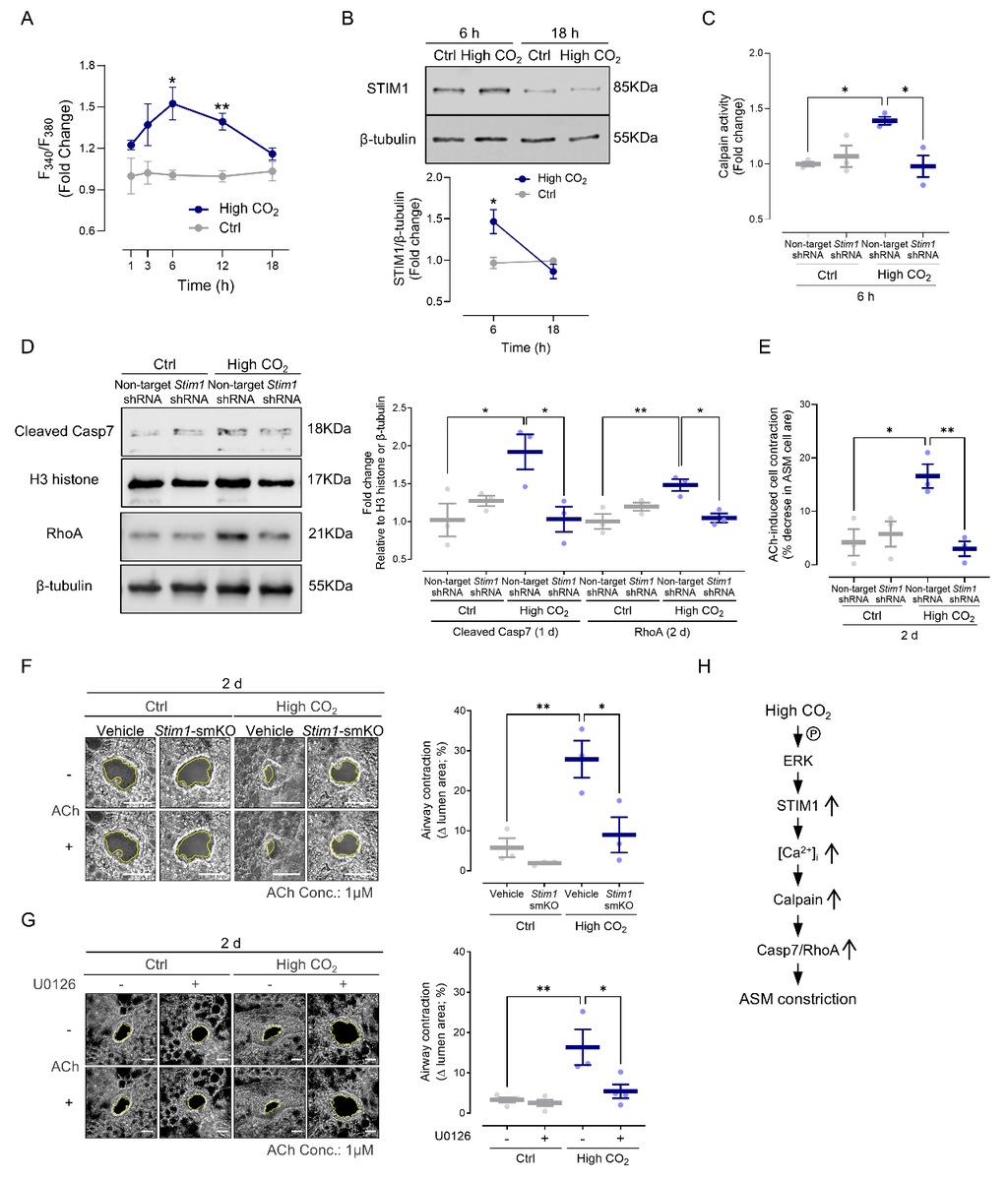
Figure 2. Blocking STIM1 protects airways: microscopy shows that airways lacking STIM1 resist constriction even under high CO2 conditions.
Human Genetic Evidence: STIM1 Variants, COPD Risk, and a Gene–Environment Interaction
Perhaps the most translational dimension of this work is the team’s integration of human genetics. Mining data from GTEx expression quantitative trait locus (eQTL) analyses, the COPD Cell Atlas single-cell RNA-seq dataset, and human case-control cohorts, the researchers uncovered a compelling narrative:
- STIM1 expression is reduced in stromal and smooth muscle cells derived from COPD patients compared to healthy controls.
- Two noncoding STIM1 variants — rs1561876 and rs3794050 — are associated with lower STIM1 expression in lung tissue and are enriched in COPD patients.
- Under normocapnic (normal CO₂) conditions, these low-expression variants correlate with lower airway resistance, consistent with a protective effect: less STIM1 means less SOCE-driven smooth muscle contraction.
- However, under hypercapnia, this protective benefit is abolished — a striking gene–environment interaction suggesting that elevated CO₂ can overcome the genetic advantage of reduced STIM1 expression, potentially by driving compensatory pathway activation or by reaching a threshold where even reduced STIM1 levels are sufficient to sustain pathologic SOCE.
This finding has immediate implications for precision medicine. It suggests that genetic stratification of COPD patients by STIM1 genotype could help identify those most vulnerable to hypercapnia-driven airway deterioration — and those who might benefit most from aggressive CO₂-lowering strategies.
Reframing Hypercapnia and Charting New Therapeutic Paths
Taken together, this study fundamentally reframes the clinical significance of hypercapnia in COPD. Rather than an inert byproduct of ventilatory failure, elevated CO₂ is an internalized environmental stressor that activates a specific, druggable signaling pathway — CO₂ → ERK → InsP3R/c-Fos → STIM1 → SOCE → sustained ASM contraction — to worsen airway obstruction. This creates a vicious cycle: airway constriction impairs ventilation, which worsens hypercapnia, which further constricts the airways.
The identification of STIM1 as the critical mechanistic node opens multiple therapeutic avenues. STIM1-targeted pharmacologic interventions, ERK pathway inhibition, and even the optimization of non-invasive ventilation strategies to reduce CO₂ burden all gain stronger mechanistic rationale from this work. The human genetic data provide a framework for identifying which patients are most likely to benefit from these approaches.
We at Precisionary Instruments are proud that the Compresstome® played a role in enabling the precision-cut lung slice assays that were essential to translating this study’s molecular findings into functional airway constriction data. The PCLS platform continues to be one of the most powerful ex vivo systems for studying airway and lung biology, and the quality of the slices is foundational to the reliability of the results.
If your research involves airway smooth muscle physiology, store-operated calcium entry, COPD pathobiology, or precision-cut lung slice functional assays, we would love to hear from you. Reach out to our team to learn how the Compresstome® vibrating microtome can support your lung research with the tissue quality your experiments demand.