Upcoming Webinar | July 7 | Investigation into Lung Immune Responses Using Precision Cut Lung Slices (PCLS)

Products
Applications
- Experiments
- Electron Microscopy
- Electrophysiology
- Genetic Sequencing (Single-Cell)
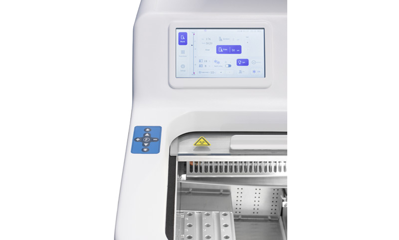
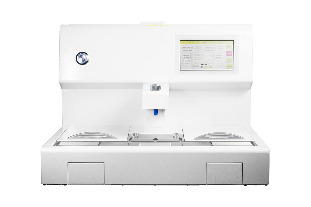
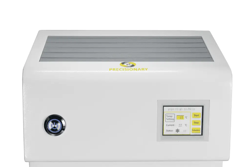
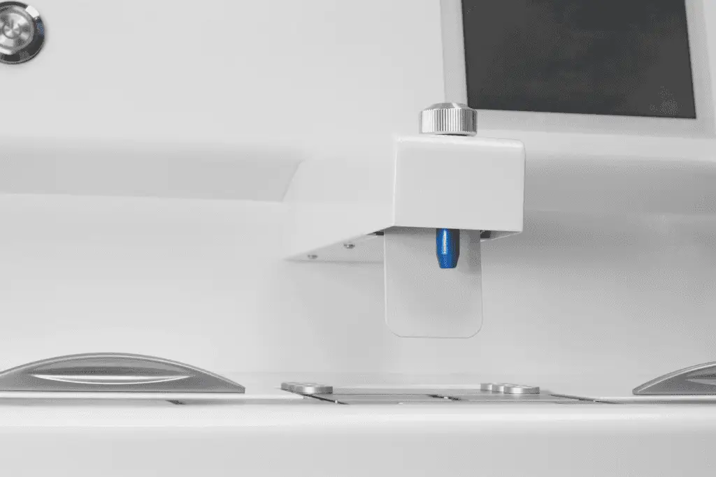
- High-throughput Sectioning
- Histopathology
- Imaging
- Immunohistochemistry
- In-situ-hybridization
- Large Sample Sectioning (Whole Organ)
- Materials & Bioengineering
- Organoids
- Organotypic Slice Culture
- Plant Research
- Precision Cut Tissue Slices
- Organs
- Brain (Fixed)
- Cartilage
- Eye
- Heart
- Liver
- Lymph-nodes
- Brain (Live or Acute)
- Embryo
- Gut (Intestines)
- Kidney
- Lung
- Tumors
- Animals
- Mouse
- Human
- Bird (Zebra Finch)
- Fish
- Frog
- Rat
- Non-Human Primates
- Chick
- Pig
E-store