Upcoming Webinar | Dec. 17 | Serial Compresstome Vibratome Sectioning for Brain Mapping

APPLICATIONS | ORGANS

Tissue Sectioning Solutions for Live Brain Slices

Create healthy, viable acute brain slices for electrophysiology with Precisionary Instruments’ Compresstome vibratomes.

High-Quality Acute Brain Slices for Electrophysiology and Calcium Imaging

Live brain tissue sectioning is critical for electrophysiology studies such as patch-clamp experiments, field recordings, and calcium imaging. These experiments require acute brain slices that remain healthy and viable for hours, ensuring the functionality of neurons throughout prolonged recordings.

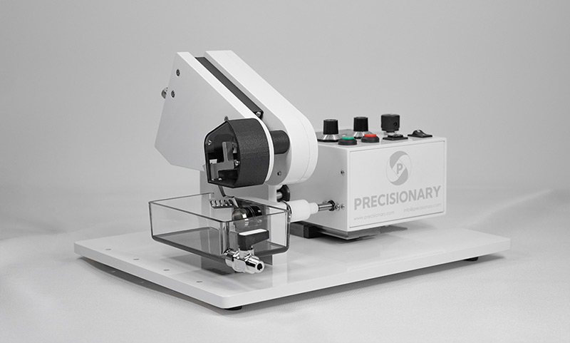
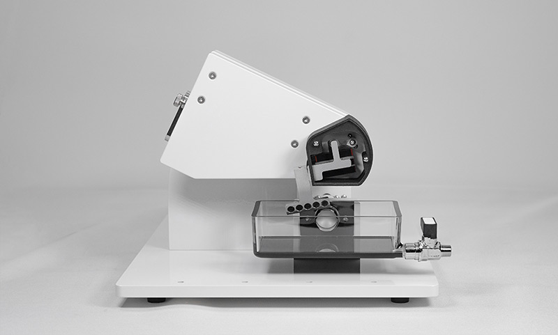
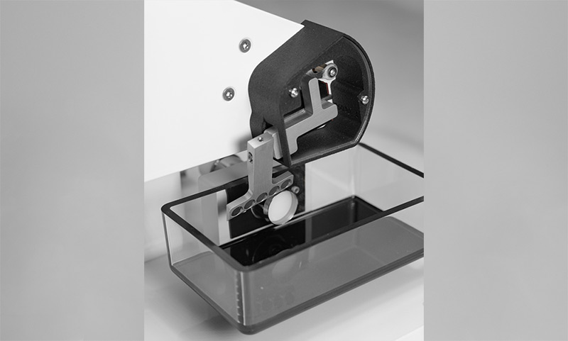
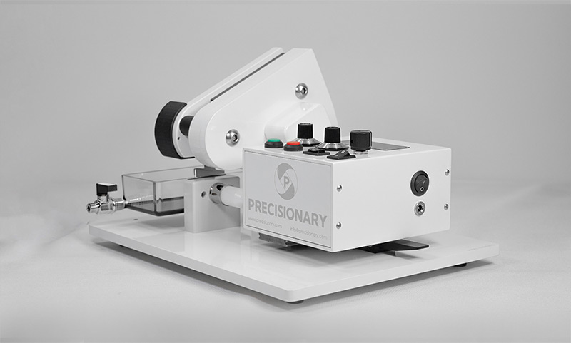
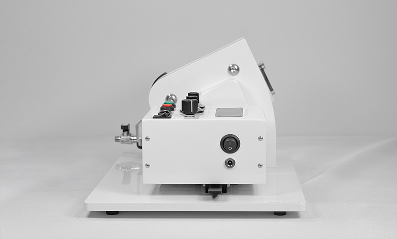
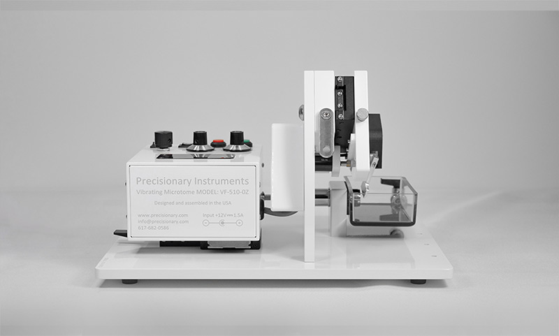
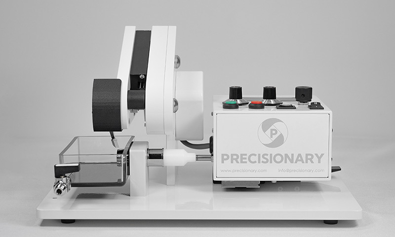
Precisionary Instruments’ Compresstome VF-510-0Z vibratome is designed to produce precise, live tissue slices that preserve cellular integrity and maximize neuron health for high-performance electrophysiology studies. Researchers can confidently use these brain slices for detailed physiological recordings, ensuring consistent, reproducible results.

Compresstome® VF-510-0Z

Precision Vibrating Microtome for Acute Brain Slices

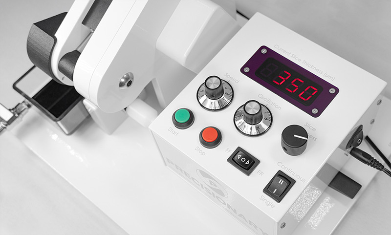
The Compresstome® VF-510-0Z vibratome is engineered to create thin, viable slices from live brain tissue, making it ideal for electrophysiology applications. This fully automated system produces healthy, consistent brain slices that retain the integrity of neurons for hours-long studies.
Experimental applications include:

With a 5-year warranty, the VF-510-0Z ensures precise, reliable tissue sectioning for neuroscience research, delivering unmatched performance in live brain tissue studies.

Real Labs Trust Precisionary Vibratomes for Their Acute Brain Slices

Reflections on a decade of patching in adult brain slices

Jonathan T. Ting is an Assistant Investigator at the Allen Institute, where he joined in 2013 to provide electrophysiology expertise for the Human Cell Types program, and to develop functional assays on human ex vivo brain slides. In this webinar, Dr. Ting discusses which key steps in the brain slice process is most important and why, and challenges our conventional beliefs of slicing solutions and methodologies.

Using electrophysiological methods to examine e-cigarette flavors’ effect on dopamine neuron function

Assistant Professor in the Department of Biomedical Sciences at Marshall University’s Joan C. Edwards School of Medicine. In addition, Dr. Henderson and his focus on the role tobacco and vaping flavors play in addiction-related behaviors, and uses the Compresstome® vibrating microtome to make all of their acute brain slices for patch-clamp electrophysiology.

Preparation of Acute Brain Slices Using an Optimized N-Methyl-D-glucamine Protective Recovery Method

Often heralded as leaders in the field, the Allen Brain institute performs pioneering research on all manner of brain tissue. Working with brain tissue can often be as frustrating as it is rewarding. Slicing brain tissue presents many challenges. The tissue is a combination of soft and fibrous regions. For over a decade, researchers at the Allen Institute for Brain Science have been using the Compresstome® vibrating microtome to help give them better brain slices with increased longevity and reduced damage to surface neurons. This enables neuroscientists to have healthy neurons for patch-clamp electrophysiology experiments.

Genetic Manipulation of the Mouse Developing Hypothalamus Through In Utero Electroporation

Researchers have used the Compresstome® in their procedure to section mouse embryo hypothalamus that has been injected with DNA and electroporated. This procedure demonstrates how it is possible to transfect nuclei in the hypothalamus region which are less accessible than those in superficial regions. Following this procedure additional experiments can be performed such as immunohistochemistry and in situ hybridization.

Investigating how sodium channels regulate the firing of cerebellar Purkinje neurons requires healthy brain slices

Dr. Ransdell explores how the Compresstome vibrating microtome is used to produce healthy brain slices for electrophysiology. He studies adult Purkinje neurons in mouse cerebellar brain slices.

Using calcium imaging and electrophysiology to explore the interaction between neurons and astrocytes in addiction

In this webinar, Dr. Wang will:

  • Discuss to quantify nascent silent synapses using electrophysiology
  • Explore how to perform ex vivo calcium imaging on brain slices
  • Share tips & tricks of brain sectioning to help reduce frustration for electrophysiology recordings

References

Booeshaghi AS, Yao Z, van Velthoven C, Smith K, Tasic B, Zeng H, Pachter L. Isoform cell-type specificity in the mouse primary motor cortex. Nature. 2021 Oct;598(7879):195-199. Epub 2021 Oct 6. PMID: 34616073; PMCID: PMC8494650. 

Driessens SLW, Galakhova AA, Heyer DB, Pieterse IJ, Wilbers R, Mertens EJ, Waleboer F, Heistek TS, Coenen L, Meijer JR, Idema S, de Witt Hamer PC, Noske DP, de Kock CPJ, Lee BR, Smith K, Ting JT, Lein ES, Mansvelder HD, Goriounova NA. Genes associated with cognitive ability and HAR show overlapping expression patterns in human cortical neuron types. Nat Commun. 2023 Jul 13;14(1):4188. PMID: 37443107; PMCID: PMC10345092. 

Egawa K, Watanabe M, Shiraishi H, Sato D, Takahashi Y, Nishio S, Fukuda A. Imbalanced expression of cation-chloride cotransporters as a potential therapeutic target in an Angelman syndrome mouse model. Sci Rep. 2023 Apr 17;13(1):5685. PMID: 37069177; PMCID: PMC10110603. 

Request a

Quote

Bring greater precision and consistency to your acute brain slices. Fill out the form to request a quote from Precisionary Instruments. We’ll respond within one business day.