

From precision-cut slices to spatial multi-omics: how modern renal research cuts and reads kidney tissue
Kidney tissue sectioning has moved well beyond the microtome and glass slide. As renal research shifts toward models that preserve living, multicellular architecture, the way we cut and maintain kidney tissue has become a scientific variable in its own right. Reproducible thickness, preserved viability, and retained cell-cell and cell-matrix interactions now determine whether a downstream fibrosis, toxicology, or spatial-omics experiment succeeds. This article surveys the current state of the art, from precision-cut kidney slices to the instrumentation that produces them, and the complementary models reshaping the field.
Precision-Cut Kidney Slices: The Centerpiece
Precision-cut kidney slices (PCKS) are among the most significant recent advances in kidney tissue sectioning. As an ex vivo model, PCKS preserve all native cell types, acellular components, and the cell-cell and cell-matrix interactions of the original tissue architecture, something dissociated cell cultures cannot reproduce (Jensen et al., 2023; Stribos et al., 2016; Stribos et al., 2016).
The technique has come a long way from the hand-cut slices of the 1920s. Modern PCKS are typically prepared at 200 to 300 µm thickness using automated slicing instruments, producing reproducible slices from both human and rodent kidneys (Poosti et al., 2015). A standardized protocol published in 2023 formalized the methodology and framed PCKS as a translational model bridging preclinical and clinical research (Jensen et al., 2023).

Figure 1. Preparation of precision-cut kidney slices. Fresh kidney tissue is harvested into ice-cold buffer, embedded in low-melt agarose, and sectioned on a vibrating microtome into 200 to 300 µm slices that remain viable in ex vivo culture for downstream histology, qPCR, viability, and imaging assays. Original schematic.
Three recent developments illustrate why PCKS have gained traction:
- Histopathological scoring for fibrosis (2026). Riishede and colleagues adapted a scoring system originally built for human kidney biopsies to quantify fibrosis and tubular atrophy in PCKS. Slices from healthy donors developed progressive fibrosis over 48 hours in culture, and TGF-β treatment intensified it. Slices from CKD patients did not accumulate fibrosis beyond baseline, suggesting the model captures disease-stage-specific biology (Riishede et al., 2026).
- Antifibrotic drug screening. TGF-β1 exposure reliably induces fibrosis markers (COL1A1, fibronectin, α-SMA), and targeted therapies such as IFNγ conjugated to PDGFRβ reduce fibrosis in this system. The multicellular environment makes PCKS a more physiologically relevant screening platform than homogeneous cell lines (Poosti et al., 2015; Stribos et al., 2016).
- Functional viability. Human PCKS remain metabolically active for more than 48 hours, maintaining glucuronidation and transporter activity. Podocyte markers such as nephrin decline during culture, which limits podocyte-specific studies but leaves tubular and interstitial biology well preserved (Stribos et al., 2016).
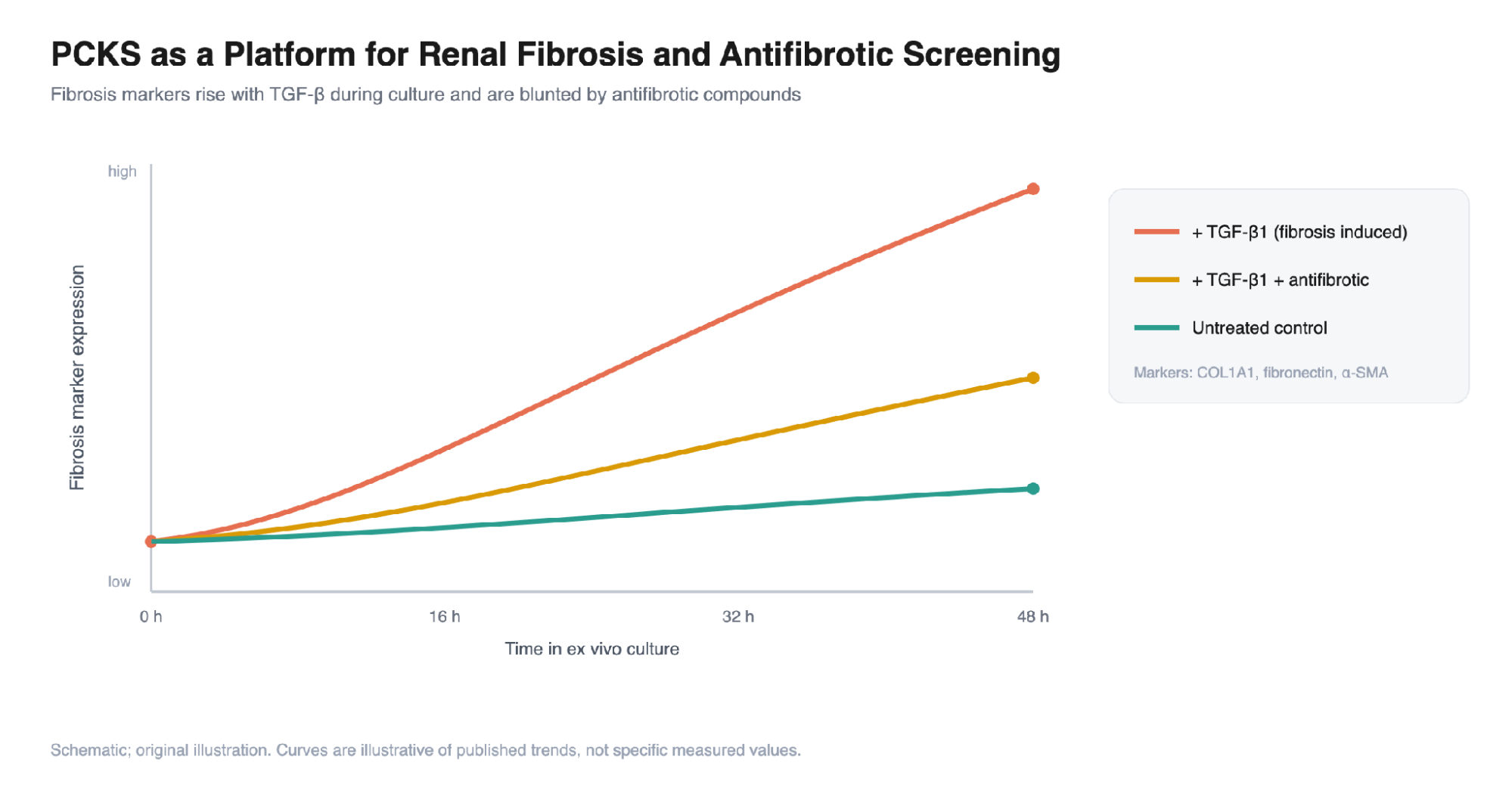
Figure 2. PCKS as a platform for renal fibrosis modeling and antifibrotic screening. During ex vivo culture, fibrosis markers rise over roughly 48 hours, are strongly amplified by TGF-β1, and are blunted when an antifibrotic compound is co-applied, while untreated slices change only modestly. Curves are illustrative of published trends, not specific measured values. Original schematic.
Instrumentation: Where Technique Becomes Reproducibility
The quality of a precision-cut slice is inseparable from the instrument that produces it. Three approaches dominate:
- Krumdieck tissue slicer. The long-standing standard for PCKS, producing consistent 200 to 300 µm slices at reasonable throughput (Poosti et al., 2015; Parrish et al., 1995).
- Vibrating-blade microtome (vibratome). Vibrating microtomes offer finer, more consistent thickness control than manual or automated slicers, and they section fresh, unfixed tissue without freezing or paraffin embedding. This is the approach best suited to viable ex vivo kidney slices, and instruments in this class have shown improved accuracy and reproducibility relative to the Krumdieck slicer across rat, mouse, and human tissue (Saitta et al., 2019; Zimmermann et al., 2009).
- Stadie-Riggs slicer. A manual option still used for ex vivo kidney preparations, particularly signaling-cascade studies, though thickness is less uniform (Saitta et al., 2019; Rao et al., 2018).

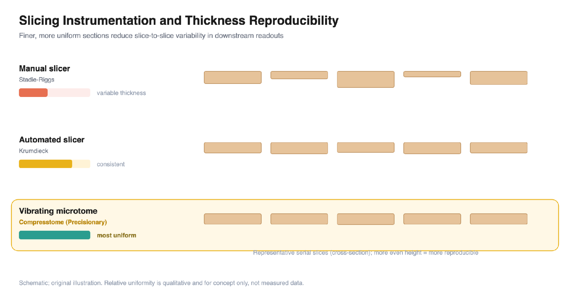
Figure 3. Slicing instrumentation and thickness reproducibility. Manual slicers (Stadie-Riggs) yield variable section thickness; automated Krumdieck slicers are more consistent; vibrating microtomes produce the most uniform serial slices. Relative uniformity is qualitative and for concept only, not measured data. Original schematic.
Vibrating-microtome workflows in kidney research
Because kidney biology depends so heavily on preserved, living architecture, the choice of instrument is not incidental. Vibrating microtomes section fresh, unfixed tissue without freezing or paraffin embedding, keeping cells viable for ex vivo culture and downstream functional assays. Precisionary’s Compresstome is purpose-built for exactly this: it pairs a vibrating blade with automated agarose embedding to deliver uniform, highly reproducible slices from soft, fresh kidney tissue, which makes it a leading choice for laboratories standardizing precision-cut kidney slice workflows and other ex vivo slice assays. It has been used in peer-reviewed kidney studies, including work on glomerular injury and the protective role of soluble Klotho (Charrin et al., 2023) and on the renal cyclooxygenase-2 / asymmetric dimethylarginine axis in mouse and human tissue (Ferreira et al., 2023). For labs that depend on slice-to-slice consistency, this combination of fresh-tissue compatibility and reproducibility is the practical foundation that the rest of the workflow depends on.
Complementary Advanced Models
PCKS do not stand alone. Several technologies address different facets of kidney complexity:
- Kidney organoids. Derived from human pluripotent stem cells, these 3D structures recapitulate nephron-like architecture with glomeruli and tubules, and model disorders from ADPKD and Alport syndrome to acute kidney injury and drug-induced nephrotoxicity. Limits remain in vascularization, developmental maturity, and reproducibility (Tekguc et al., 2022; Xi & Song, 2025).
- Kidney-on-a-chip. Microfluidic platforms reconstruct individual nephron segments under perfusion. Fluid shear stress improves proximal tubule phenotype and transporter expression, and primary human proximal tubule cells can stay viable and polarized for up to six months. A 2026 review highlighted integration of hiPSC-derived organoids with real-time biosensing, 3D bioprinting, and AI-guided automation (Huang et al., 2024; Ma et al., 2026; Nieskens & Wilmer, 2016; Chalker et al., 2025).
- Organoid-on-chip hybrids. Aceves and colleagues built a proximal tubule-on-chip from organoid-derived epithelial cells, with upregulated OCT2 and OAT1/3 transporters and better nephrotoxicity prediction than immortalized lines (Aceves et al., 2022).
Spatial Transcriptomics and Multi-Omics
A parallel revolution is happening not in how tissue is cut, but in what we read from the section once it is on the slide.
- The Kidney Precision Medicine Project (KPMP) built an integrated reference atlas using single-cell and single-nucleus transcriptomics, laser microdissection, CODEX imaging, and spatial metabolomics across 56 subjects (Hansen et al., 2022).
- A 2023 Nature atlas applied multiple single-cell assays (more than 400,000 nuclei and cells) and spatial imaging to 93 kidneys, defining 51 main cell types and 28 cellular states altered in injury (Lake et al., 2023).
- Single-cell multi-omic and spatial profiling has resolved four kidney microenvironments (glomerular, immune, tubule, fibrotic), with the fibrotic microenvironment outperforming traditional histopathology for prognosis (Abedini et al., 2024).
For anyone sectioning kidney tissue today, these methods raise the bar: section quality and preservation now determine the ceiling on spatial-omics resolution.
Limitations and Where the Field Is Headed
No current model is complete. PCKS lack blood flow, osmotic gradients, and immune-cell trafficking, limiting their fidelity to in vivo physiology (Riishede et al., 2026). Organoids remain immature and poorly vascularized (Xi & Song, 2025). Bioengineering strategies are converging on these gaps: endothelial co-culture, xenografting onto the chick chorioallantoic membrane for vascularization, and integration with microfluidic platforms (Kaisto et al., 2020; Ma et al., 2026).
The most promising frontier is convergence. Pairing precision-cut kidney slices, which preserve native tissue architecture, with spatial transcriptomics and multi-omics readouts offers a path to mechanistic insight that neither approach reaches alone. As instrumentation continues to improve the reproducibility of the cut itself, the section on the slide becomes a more faithful window into kidney biology and disease.
Ready to bring precision to your kidney sectioning?
From establishing a precision-cut kidney slice protocol to scaling an ex vivo drug-screening program, the Precisionary team can help you choose the right instrument and dial in a reproducible workflow for your tissue. The Compresstome vibrating microtome is trusted in peer-reviewed kidney research for viable, uniform slices from fresh, unfixed tissue.
→ Explore the Compresstome vibrating microtomes
→ See kidney sectioning applications (PCKS)
→ Talk to our team: Contact Us
Email [email protected] Call 617-682-0586