Wildenberg Lab Supported by Precisionary Instruments
In our latest Lab Spotlight, we’re thrilled to feature the groundbreaking work of Dr. Gregg Wildenberg and his team at The University of Chicago. Dr. Wildenberg also holds a joint appointment at Argonne National Laboratory. Dr. Wildenberg’s lab, composed of a dynamic team of eight individuals, is dedicated to unraveling the mysteries of neuroanatomy using cutting-edge technology and innovative approaches.

Exploring Scalable Neuroanatomy
At the heart of Dr. Wildenberg’s research lies a profound interest in uncovering the organizational principles of the brain. Leveraging sophisticated equipment including scanning electron microscopes and Argonne’s synchrotron source X-ray computed tomography, the lab delves into scalable neuroanatomy, aiming to shed light on the intricate workings of the brain.
Compresstome Vibratome: Illuminating Neuroanatomy
One instrumental tool in Dr. Wildenberg’s arsenal is the Compresstome, a vibratome designed and manufactured by Precisionary Instruments. In a recent series of experiments, Dr. Wildenberg and his team utilized the Compresstome to trace myelinated axons across serial brain sections. By staining the sections with heavy metals, they were able to label all myelinated axons, enabling detailed analysis using electron microscopy and X-ray imaging.
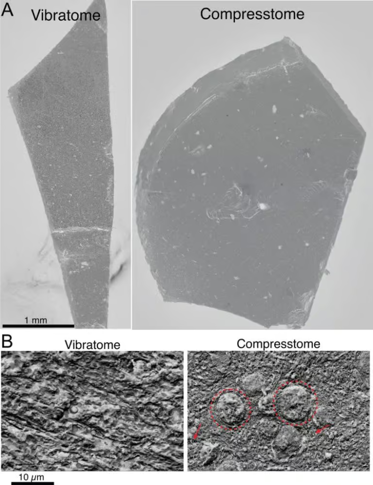
The results were striking. Despite the rigorous sectioning process, Dr. Wildenberg observed minimal tissue loss between Compresstome sections, allowing for seamless tracing of myelinated axons. With a calculated tissue loss of approximately 700nm, the Compresstome demonstrated unparalleled precision in sectioning. Furthermore, imaging of the section surfaces revealed distinct advantages of the Compresstome over traditional vibratomes, with biological features clearly visible on Compresstome sections.

To observe the visual evidence supporting Dr. Wildenberg’s findings, the team analyzed SEM images of brain tissue sections. Figure A above displays a low-resolution comparison between sections obtained using a vibratome (left) and the Compresstome (right). In Figure B, high-resolution SEM images reveal distinct differences in surface quality. The vibratome section (left) appears jagged, lacking identifiable biological features. In contrast, the Compresstome section (right) exhibits clear visualization of neuronal nuclei (dotted circle) and individual myelinated axons (MAs), as indicated by red arrows. This retention of biological architecture on the cut section’s surface highlights the Compresstome’s precision and fidelity in neuroanatomy research. Scale bars of 1 mm (Figure A) and 10 µm (Figure B) provide further context for the detailed examination of tissue structure.
Compresstome Vibratome Enables Cutting-Edge Neuroanatomy Research
As the makers of the patented Compresstome, Precisionary Instruments is proud to play a pivotal role in Dr. Wildenberg’s groundbreaking research. By providing a tool that offers exceptional precision and minimal tissue loss, the Compresstome empowers researchers to explore the complexities of neuroanatomy with unprecedented detail and accuracy.
With ongoing advancements in technology and a relentless pursuit of scientific discovery, Dr. Wildenberg and his team continue to push the boundaries of neuroanatomy research. Through their innovative approaches and collaborative spirit, they are poised to unlock new insights into the inner workings of the brain, shaping the future of neuroscience.
Precisionary Instruments is honored to partner with Dr. Gregg Wildenberg and his team, supporting their mission to unravel the mysteries of the brain and advance our understanding of neuroanatomy.
Interested in featuring your lab in our Lab Spotlight series? Contact us today to share your innovative research and explore how our instruments can enhance your scientific endeavors.
Stay tuned for more inspiring stories from the forefront of scientific discovery.
