

The human brain, a complex network of billions of neurons, remains one of the greatest enigmas of science. In the quest to decode its mysteries, scientists have embarked on an ambitious endeavor called “projectomes.” Projectomes are comprehensive maps of the brain that detail the trajectories of individual long-distance projecting neurons, shedding light on how different brain regions communicate with one another. The latest publication, authored by Dr. Gregg Wildenberg and his colleagues from the University of Chicago and Argonne National Library, is a monumental step forward in this pioneering field.
Projectome Project Pipeline
In their publication titled “A Pipeline for a Primate Projectome: Mapping Every Individual Myelinated Axon Across the Whole Brain,” Dr. Wildenberg and his team introduce a groundbreaking approach to neuroscience. The research involves mapping the entire trajectory of every long-distance projecting neuron in the non-human primate brain, essentially creating a comprehensive projectome. Here’s how it works in a nutshell (Figure 1):

Figure 1. Proposed pipeline for the projectome tracing study. Step 1: Slicing into Pillars. Imagine the brain as a puzzle. Scientists cut it into manageable pieces, like slices of cake that are further divided into smaller “pillars. Step 2: Staining and X-ray Imaging. These pillars are treated with heavy metals and imaged using synchrotron X-ray technology, revealing their intricate structure. Step 3: Tracing Axons. Researchers trace individual myelinated axons (MAs), the brain’s information highways, within and across each pillar, creating a communication map. Step 4: Exploring Synapses. The same pillars are then used for electron microscopy, allowing scientists to explore the brain’s microscopic synaptic connections.
This breakthrough reveals the brain’s inner workings and holds great promise for understanding neurological disorders and brain function. The brain’s mysteries are gradually unfolding, thanks to projectome mapping and advanced imaging.
Major Findings From The Projectome Study
In the publication, the team share the following key findings:
- Compresstome Vibratome’s Role: The study also explored the use of the ‘Compresstome,’ a microtome designed to compress tissue during cutting. The Compresstome proved to be highly effective in tracing individual MAs across tissue sections, offering superior performance compared to other vibratomes tested. The surfaces of Compresstome-cut brain sections were relatively preserved, making it easier to identify biological features like soma, nuclei, and individual MAs, while standard vibratome sections exhibited uniformly jagged surfaces without discernible biological features.
- Mapping the Brain: The study presents an innovative experimental pipeline designed to map individual myelinated axons (MAs) and cell morphologies across large brains using synchrotron source X-ray imaging.
- Addressing Challenges: The research team successfully addressed several critical challenges, including optimizing methods for brain sectioning with minimal tissue loss, determining the minimal resolution required for tracing individual MAs within volumes and across tissue gaps, automating the segmentation of individual MAs, and developing algorithms for joining segments across tissue gaps.
- High Tracing Accuracy: With current X-ray resolutions (100 nm voxels), the study achieved remarkable accuracy in tracing MAs. They reported near 100% accuracy within pillars and 97% human accuracy and 94% algorithmic accuracy in tracing MAs across gaps estimated to be 2-fold wider than the actual gap between Compresstome-cut sections.
- Success Rate Model: The researchers introduced a predictive model for tracing success rate, which takes into account factors like the fraction of axons within a pillar that are reconstructed without error, the fraction of axons successfully traced across a kerf gap, and the number of gaps jumped by the myelinated axon from end to end. This model suggests that algorithms could potentially trace the long-distance MA projections of 10% of all MAs in a cubic centimeter volume.
These findings represent significant advancements in the field of neuroscience and connectomics, offering researchers valuable tools and insights for mapping the intricate neural networks of the brain with unprecedented precision and efficiency.
The Role of the Compresstome Vibratome
One critical aspect that has enabled the success of this project is the utilization of our company’s Compresstome vibratome. Designed to minimize tissue damage during the cutting of living brain slices, the Compresstome proved to be a game-changer for the projectome project. The research team tested various vibratomes, but it was the Compresstome that emerged as the superior choice.
The Compresstome demonstrated its capability to preserve the integrity of brain sections, allowing for the identification of individual myelinated axons across cut surfaces. In contrast, standard vibratome sections exhibited jagged surfaces devoid of any recognizable biological features. This crucial advantage of the Compresstome not only facilitated the tracing of myelinated axons but also contributed significantly to the success of the projectome project as a whole.
Seeing is believing, so here are some visual results in Dr. Wildenberg’s publication:
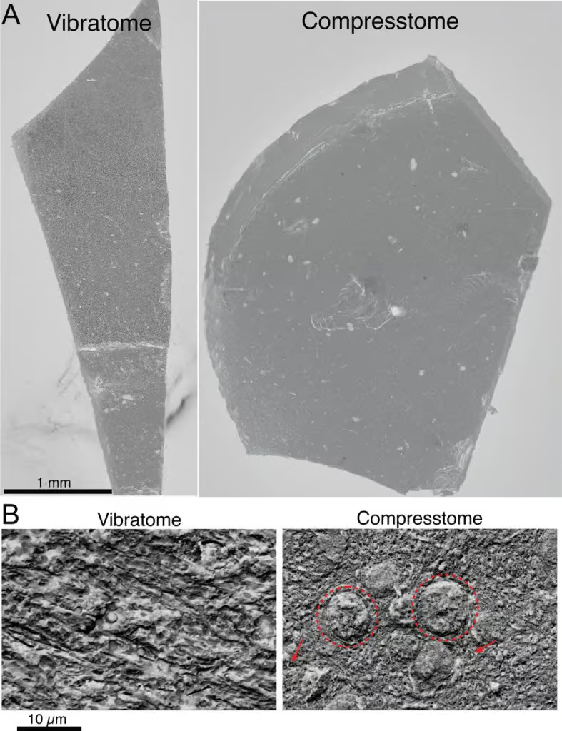
Figure 2. Understanding the Compresstome’s Impact on Brain Tissue. A. In the images on the left (vibratome) and right (Compresstome), we see brain tissue that has been cut into slices. B. When we zoom in closer, we notice something remarkable. In the vibratome section (left), it’s challenging to see the details of the brain’s structure. However, in the Compresstome section (right), we can clearly see the nuclei of brain cells (shown with dotted circles) and individual myelinated axons (highlighted with red arrows). This means that the Compresstome preserves the fine details of the brain’s biological architecture on the very surface of the cut section.
Implications for Your Research
As a scientist or researcher, you might be wondering how the findings of this groundbreaking publication can impact your own work. To answer this question, let’s turn to a quote from Dr. Jian-Qiang Kong, the President of Precisionary Instruments, the company behind the Compresstome. Dr. Kong stated, “This research overcame several crucial obstacles and technical challenges, theoretically proving that the Projectome project on a non-human primate brain is both feasible and reliable.” He goes on to highlight that the Compresstome played a pivotal role in solving the critical issue of tracing myelinated axons across sections, ultimately completing a vital piece of the Projectome puzzle.
The implications of this research are profound. It opens doors to a deeper understanding of brain microstructure, biological function, and the mechanisms underlying neurological disorders. The Compresstome, with its unique capabilities, has proven to be an indispensable tool for researchers in the field of neuroscience. As you embark on your own investigations into the mysteries of the brain, you can rest assured that this pioneering work has paved the way for more discoveries and advancements in the realm of brain science.