

Key Takeaways
- Byun et al. designed a fully 3D-printed recording chamber system for acute brain slice electrophysiology — a submerged chamber, tissue-positioning stage, and suction assembly — built for roughly $1.23 in material cost versus the thousands of dollars commercial systems typically require.
- Thermal imaging and thermistor testing confirmed uniform perfusate temperature distribution and a stable 36°C bath setpoint, ruling out the temperature gradients that commonly degrade recording quality in poorly designed chambers.
- The platform produced high-fidelity fEPSP recordings from mouse hippocampal CA1 slices, with graded input-output curves and stable baselines over a 15-minute recording window — performance on par with commercial recording chambers.
- Hippocampal slices were prepared on a Compresstome vibrating microtome, preserving intact superficial dendrites and minimizing compression artifact so slice quality wouldn’t confound validation of the new chamber design.
- All design files are released open-source under CERN-OHL-S-2.0 and CC BY 4.0, letting any lab with a basic FDM 3D printer reproduce the platform.
Access to cutting-edge neuroscience tools shouldn’t depend on a lab’s budget. That’s the driving philosophy behind a new study from Younsoo Byun and colleagues, published in HardwareX: “A low-cost, 3D-printed open-source platform for acute brain slice electrophysiology”. In this work, Byun et al. (2026) designed, fabricated, and validated a fully integrated recording chamber system — built entirely from 3D-printed PLA at a material cost of roughly $1.23 — and demonstrated that it can produce high-fidelity field excitatory postsynaptic potential (fEPSP) recordings from acute mouse hippocampal slices. The achievement is a meaningful step toward making electrophysiology accessible to resource-limited and early-stage neuroscience laboratories around the world.
The Price Tag Problem in Electrophysiology
Acute brain slice electrophysiology is one of the most powerful techniques in neuroscience. It allows researchers to study synaptic transmission, network dynamics, and pharmacological responses in living neural circuits with remarkable precision. But there’s a catch: the hardware required to do it — submerged recording chambers, perfusion systems, tissue-positioning stages, and suction assemblies — often costs several thousand dollars from commercial suppliers. For a well-funded lab at a major research university, that expense is manageable. For labs at teaching-focused institutions, in low- and middle-income countries, or for graduate programs just getting started, these costs can be prohibitive.
The result is an uneven playing field. Talented neuroscientists at under-resourced institutions may simply lack access to the instrumentation needed to train students in electrophysiology or to pursue their own research questions. Byun and colleagues set out to change that by asking a straightforward question: can you replicate the functional requirements of a commercial submerged recording chamber using nothing more than a consumer-grade FDM 3D printer and standard PLA filament?
A Three-Module System for Under Two Dollars
The team’s answer is an elegantly modular platform consisting of three independently printed and assembled components: a submerged recording chamber, a tissue-positioning stage, and a suction assembly. The entire system uses approximately 22.45 grams of PLA filament, bringing the total material cost to about 1,749.88 KRW — roughly $1.23 USD. No specialized tools, adhesives, or post-processing steps are required for assembly. Each module can be independently replaced without rebuilding the entire system, and the platform is designed to integrate seamlessly with standard commercial perfusion, heating, and electrophysiology hardware that labs may already own.
Crucially, the team didn’t stop at fabrication. They subjected the platform to rigorous thermal and electrophysiological validation to ensure it could meet the demands of real experimental work.
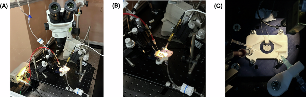
Figure 1. The complete 3D-printed recording setup in action on the microscope rig, ready for brain slice experiments.
Thermal Stability and Electrophysiological Performance in the Hippocampal Slice
Reliable brain slice recordings depend on precise, uniform temperature control. Neurons are exquisitely sensitive to thermal fluctuations, and uneven perfusate distribution can create temperature gradients that compromise data quality. The researchers addressed this concern with two complementary approaches:
- Infrared thermal imaging demonstrated that heated perfusate dispersed uniformly across the chamber bath within three minutes, with no stagnant peripheral zones — a common issue in poorly designed chambers that can lead to localized cell death or inconsistent synaptic responses.
- Thermistor-based measurements confirmed that bath temperature reached the 36°C target setpoint within approximately three minutes and remained stable over a nine-minute observation period, with only a transient initial overshoot before settling.
With thermal performance validated, the team turned to the ultimate test: could the chamber support high-quality electrophysiological recordings? Using acute hippocampal slices prepared from adult male C57BL/6N mice, they recorded fEPSPs from the CA1 stratum radiatum following stimulation of the Schaffer collateral pathway — a gold-standard preparation in synaptic physiology. The results were convincing:
- Input-output curves showed a graded, monotonic stimulus-response relationship across stimulation intensities from 0 to 600 µA, consistent with the expected progressive recruitment of synaptic inputs.
- Baseline fEPSP amplitudes remained stable over a 15-minute recording period with minimal inter-sweep variability, indicating that the platform can support the kind of extended baseline recordings required for long-term potentiation (LTP) and long-term depression (LTD) experiments.
- Signal fidelity was high throughout, with clean waveforms that reflect well-maintained slice health during recording.
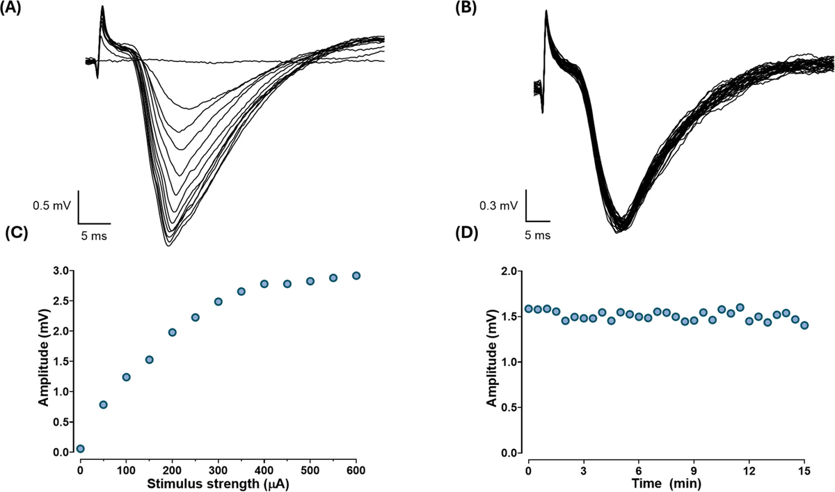
Figure 2. Electrical recordings show strong, stable brain signals — proof that this budget-friendly device performs like standard lab equipment.
Starting with a Clean Slice: How the Compresstome Enabled This Work
Even the best recording chamber in the world can’t rescue a poorly prepared brain slice. The quality of electrophysiological data begins at the moment of sectioning, and this is where the researchers’ choice of tissue preparation instrument mattered. Byun et al. used the Precisionary Instruments Compresstome® VF-210-0Z vibrating microtome to prepare 350 µm coronal hippocampal slices from their C57BL/6N mouse brains.
The Compresstome’s agarose embedding and compression technology was particularly well-suited to this study’s goals. In acute brain slice electrophysiology, the health of the superficial cell layers is critical — these are the neurons closest to the recording and stimulating electrodes. Traditional vibratomes can cause compression artifacts and mechanical damage to the tissue surface during cutting, leaving a layer of dead or damaged cells that degrades signal quality and forces researchers to record from deeper, less accessible regions.
The Compresstome addresses this by stabilizing the tissue within an agarose block that is gently advanced through a compression tube during sectioning. This approach minimizes the lateral displacement and vibration-induced tearing that plague conventional vibratomes, producing slices with cleaner, healthier surfaces. For Byun and colleagues, this meant that the hippocampal slices placed into their 3D-printed chamber had the best possible starting condition — intact superficial dendrites in the CA1 stratum radiatum, healthy Schaffer collateral axon terminals, and minimal compression artifact. The stable baselines and clean input-output curves they recorded are a direct reflection of both the chamber’s performance and the quality of the slices that went into it.
In a study designed to validate a novel, low-cost recording platform, controlling for slice quality was essential. Had the researchers used a sectioning method that introduced variability in tissue integrity, it would have been impossible to determine whether any signal issues originated from the chamber design or from compromised tissue. The Compresstome provided that critical control.
Opening Doors for Neuroscience Worldwide
Perhaps the most impactful aspect of this work is the team’s commitment to open science. All design files have been released under the CERN Open Hardware Licence (CERN-OHL-S-2.0), and all documentation is available under a Creative Commons CC BY 4.0 license. Any lab with access to a basic FDM 3D printer can download the files and produce a functional recording chamber. The modular design means components can be customized for specific experimental needs or replaced individually if damaged — no need to reorder an entire commercial system.
It’s worth noting the current limitations honestly, as the authors do: the platform is validated for extracellular field recordings and is not currently compatible with infrared differential interference contrast (IR-DIC) visualization needed for visually guided patch-clamp electrophysiology. But for the substantial body of neuroscience research that relies on field potential recordings — LTP/LTD studies, pharmacological screening, network oscillation analysis, and educational training — this platform offers a viable, validated, and remarkably affordable alternative to commercial systems.
This study stands as a compelling example of how thoughtful engineering, open-source principles, and rigorous validation can expand access to fundamental neuroscience techniques. By pairing innovative chamber design with high-quality tissue preparation using the Compresstome, Byun et al. have shown that budget constraints don’t have to mean compromised science.
If your lab is working on acute brain slice electrophysiology — whether you’re building out a new rig, equipping a teaching laboratory, or looking for ways to improve slice quality in your existing workflow — we’d love to hear from you. Reach out to us at precisionary.com to learn how the Compresstome vibrating microtome can support your electrophysiology research.