


At Precisionary Instruments, our ultimate goal is to support scientists throughout their careers and we absolutely love following them as they become successful in their respective fields. There is nothing more rewarding than partnering with brilliant minds, watching their projects evolve, and celebrating their monumental milestones. Today, we are thrilled to shine a Lab Spotlight on a recent publication from Dr. Joannah Fergusson.
Dr. Fergusson is a Scientific Officer within the IAT Theme at the NIHR Biomedical Research Centre, Kennedy Institute for Rheumatology, at the University of Oxford.
A Valued History of Collaboration
Our relationship with Dr. Fergusson has been an ongoing highlight for our team. She previously hosted an incredibly insightful webinar exploring how to investigate vaccine adjuvant-induced inflammation using precision-cut slices, and we are eagerly hoping she will join us again as a guest speaker very soon.
Beyond sharing her expertise with our community, Dr. Fergusson has been a dedicated collaborator. Late last year, she provided a key letter of support for our MLSC Bits to Bytes grant proposal. Our proposed project focuses on developing best practice protocols for precision-cut tissue slices (PCTS) and generating well-annotated multiomics reference data for PCTS in culture conditions. Her endorsement strongly reinforced our proposal, highlighting a shared vision as her team embarks on complementary standardization studies with lymphoid tissues.
Recent Publication in STAR Protocols
We are ecstatic to share that Dr. Fergusson has recently had a paper accepted and officially published in STAR Protocols.
The paper is titled: “Protocol for precision cutting and short-term culture of lymph node tissue slices for modeling of innate immune responses ex vivo”.
You can read her full, officially published protocol here: https://doi.org/10.1016/j.xpro.2026.104421.
Summary of the Publication & Why It Matters
In her newly published work, Dr. Fergusson presents a protocol that enables the highly organized microanatomical structure of secondary lymphoid organs to be preserved during short-term culture. This expands on 2D cell culture and organoid approaches by maintaining the native 3D lymphoid microarchitecture, which allows for physiologically relevant insights into immune perturbation.
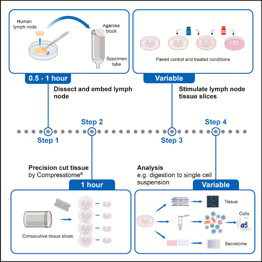
Here is a closer look at the methodology and its significance:
- Direct Human Tissue Use: The protocol utilizes human cystic lymph nodes obtained as a by-product of routine cholecystectomy surgeries.
- Addressing Anatomical Challenges: Human lymph nodes are larger, more elastic, and significantly more difficult to cut compared to mouse lymph nodes. This optimized protocol provides guidance to specifically overcome these hurdles.
- Live Tissue Cross-Sections: The paper details the steps for precision cutting human lymph nodes into live tissue cross-sections. Utilizing cross-sections of the full organ enables a broad representation of lymph node substructures in a standardized manner.
- Short-Term Culture with Stimuli: These tissue slices can be successfully cultured for up to 20 hours alongside innate immune stimuli.
- Comprehensive Multi-Readout Analysis: The cultured slices can be evaluated using multiple downstream readouts. These include secretome analysis, immunohistochemistry, and digestion into single-cell suspensions for transcriptomics or flow cytometry.
- Paired Condition Testing: Because multiple tissue slices can be generated from a single donor, researchers can perform paired analyses of both treated and untreated conditions, which is crucial for managing donor-to-donor variation.
Ultimately, this methodology provides researchers with a robust, comprehensive ex vivo model for mechanistic studies of human lymphoid tissue and innate immune responses.
How the Compresstome Made It Possible
A core requirement for establishing this reliable ex vivo model is the ability to carefully slice live, soft human lymph nodes. Dr. Fergusson utilized the Compresstome® VF-510-OZ to achieve these precision-cut human lymph nodes.
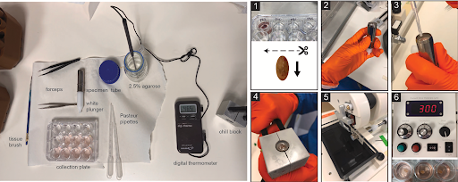
Figure 3. Overview of components required for tissue embedding (left) and the embedding and cutting procedure (1–6)
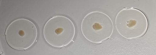
According to the protocol, the lymph nodes are embedded in 2.5% agarose , and cutting at thicknesses of 300-400 µm generates consistent cross-sections from the live tissue. Because the Compresstome effectively stabilizes the delicate tissue during the cutting process, it yields highly viable slices capable of thriving in short-term culture, making this groundbreaking functional model a reality.
To learn more about optimizing your own lymphoid tissue sectioning, please visit our Lymph Nodes Application Page.
Learn More & Get Connected
- Explore her work: Visit the Dr. Joannah Fergusson Profile Webpage to dive deeper into her ongoing research.
- Join the conversation: If you’re interested in finding out more about Dr. Fergusson’s work, let us know and we can connect you!
- Equip your lab: Do you need a Compresstome vibratome to get your research accomplished? Ask us!