

Key Takeaways
- IL-33 increased airway contraction and blunted the relaxation response to the rescue bronchodilator formoterol in non-diseased human lung tissue, suggesting it can impair bronchodilator effectiveness independently of inflammation.
- IL-25 selectively increased airway contraction in asthma-derived tissue but had little effect in non-diseased tissue, pointing to a disease-specific vulnerability.
- TSLP did not significantly alter airway contraction or relaxation in either tissue type, demonstrating that the three alarmins are not interchangeable in their effects on airway tone.
- Human precision-cut lung slices (hPCLS) prepared with the Compresstome vibrating microtome enabled the researchers to study small airway contraction and relaxation ex vivo with intact tissue architecture, preserving the functional responses needed to detect these subtle pharmacological differences.
- These findings could inform how clinicians match anti-alarmin biologic therapies to specific asthma phenotypes, targeting not just inflammation but direct effects on airway smooth muscle tone.
When a person with asthma reaches for a rescue inhaler, the expectation is rapid relief: airways relax, breathing eases. But what if certain immune signals are actively working against that bronchodilator, tightening the airway in ways that inflammation alone doesn’t explain? A new study published in Respiratory Research by Kennedy et al. (2026) digs into exactly this question, revealing that three alarmins, TSLP, IL-33, and IL-25, exert strikingly different effects on airway contraction and relaxation in human small airways, and that those effects depend on whether the tissue comes from a non-diseased or asthma-affected donor.
The work was led by Joshua L. Kennedy, Dana N. Frederick, Reynold A. Panettieri Jr., and Cynthia J. Koziol-White, and it challenges the assumption that alarmins influence asthma primarily through their well-characterized inflammatory pathways. Instead, the team provides evidence that some alarmins can directly modulate bronchomotor tone, the balance of contraction and relaxation that determines how open or closed an airway is at any given moment.
The Missing Piece: Airway Tone Beyond Inflammation
Alarmins are epithelial-derived cytokines released in response to allergens, viruses, pollutants, and other insults. TSLP, IL-33, and IL-25 sit at the very top of the inflammatory cascade in asthma, and all three are now therapeutic targets: anti-TSLP (tezepelumab) is already approved, and anti-IL-33 and anti-IL-25 agents are in clinical development. The clinical rationale has been built largely on these molecules’ ability to amplify type 2 inflammation: recruiting eosinophils, activating mast cells, and promoting mucus production.
But asthma is fundamentally a disease of airway obstruction, and obstruction is driven not only by inflammation but also by smooth muscle contraction and impaired relaxation. Whether alarmins contribute to that mechanical dysfunction directly, separate from their inflammatory signaling, has been far less studied. Kennedy and colleagues set out to fill that gap by asking a deceptively simple question: if you expose a living human airway to each alarmin individually, does the airway contract more tightly and relax less readily?
IL-33 Tightens Airways and Blunts Bronchodilator Rescue
Using human precision-cut lung slices (hPCLS) from deidentified non-diseased donor lungs, the researchers measured airway lumen area by video microscopy while challenging the tissue with the contractile agonist carbachol and then relaxing it with the beta-agonist formoterol, the same class of drug found in rescue inhalers. Dose-response curves and area-under-the-curve (AUC) analyses allowed them to quantify both the magnitude and sensitivity of each response.
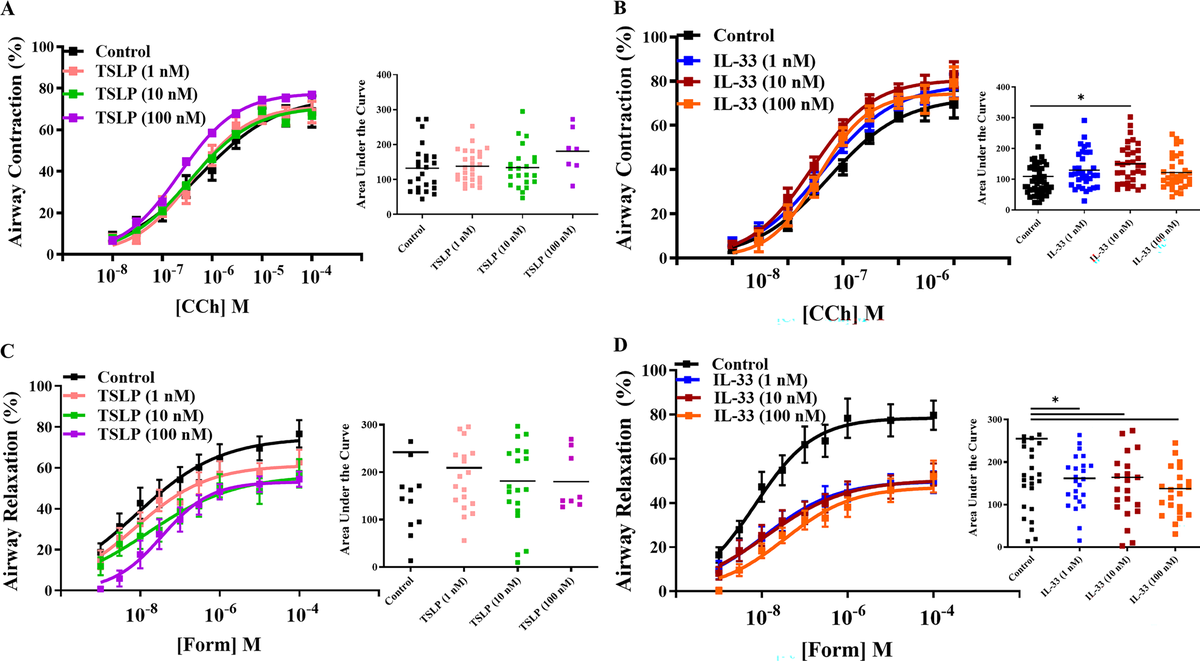
Figure 1. IL-33, but not TSLP, makes small airways contract more strongly and relax less effectively in response to bronchodilator treatment. Source: Kennedy et al., Respiratory Research (2026).
The results drew a clear line between IL-33 and TSLP. IL-33 treatment significantly increased carbachol-induced contraction in non-diseased airways, meaning the same dose of a contractile stimulus produced a more exaggerated narrowing of the airway lumen. Even more concerning, IL-33 reduced the relaxation response to formoterol. In practical terms, this means an airway exposed to IL-33 not only squeezes tighter but also responds less well to the very medication a patient would use for acute relief. TSLP, by contrast, had no significant effect on either contraction or relaxation in the same tissue.
This finding is particularly provocative because it suggests IL-33 could be undermining bronchodilator efficacy in real time, through a mechanism that operates alongside, but independently of, its role in driving eosinophilic inflammation. For patients whose asthma features high IL-33 activity, poor rescue inhaler response might not just be a matter of severe inflammation; IL-33 may be directly stiffening the airway smooth muscle.
IL-25’s Selective Grip on Asthma-Derived Airways
The second arm of the study compared the effects of IL-25 and TSLP on carbachol-induced contraction in both non-diseased and asthma-derived donor tissue. Here, the disease context proved decisive.

Figure 2. IL-25 boosts airway muscle contraction in asthma-derived tissue, but has little effect on healthy airways, unlike TSLP, which shows no such difference. Source: Kennedy et al., Respiratory Research (2026).
IL-25 significantly increased airway contraction specifically in asthma-derived tissue while producing little effect in non-diseased airways. TSLP, once again, showed no significant contractile effect in either group. This disease-specific sensitivity to IL-25 suggests that something about the asthmatic airway, whether it is receptor upregulation, altered downstream signaling, or structural remodeling, primes it to respond to IL-25 with heightened contraction. Non-diseased tissue appears largely indifferent to the same signal.
The implication is striking: IL-25 may be a driver of hypercontractility that only matters once the airway has already been remodeled by disease. This kind of context-dependent effect would be invisible in studies using only healthy tissue and underscores the value of studying asthma-derived specimens directly.
Precision-Cut Lung Slices and the Role of Uniform Sectioning
This study’s ability to detect subtle, alarmin-specific differences in contraction and relaxation depended critically on the quality and consistency of the lung slices themselves. The researchers prepared hPCLS using the Compresstome oscillating vibratome, which employs an agarose-compression approach to stabilize the soft, spongy lung tissue during sectioning.
Human lung parenchyma is notoriously difficult to section uniformly. The tissue is porous and compliant, and without adequate stabilization, a vibrating blade can compress or tear the delicate small airways rather than cutting cleanly through them. The Compresstome addresses this by embedding the tissue in an agarose block that is advanced through a compression tube, holding the specimen firmly in place so the oscillating blade can produce slices of consistent thickness with minimal mechanical artifact.
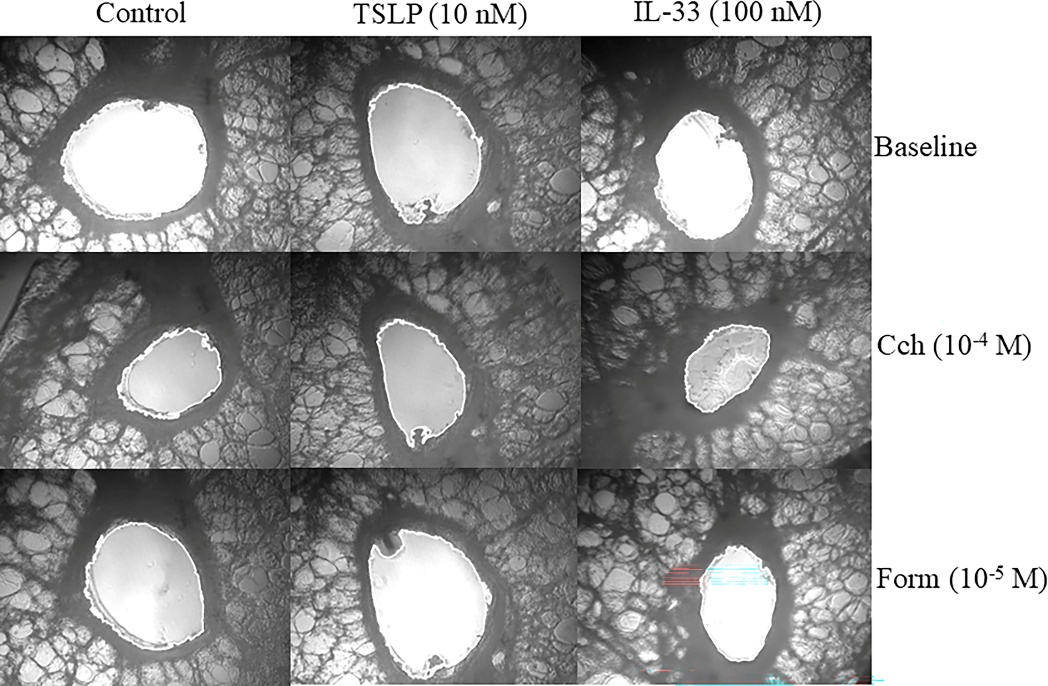
Figure 3. A representative airway within a Compresstome-prepared hPCLS, imaged at baseline, after carbachol-induced contraction, and after formoterol-induced relaxation, across control, TSLP, and IL-33 conditions. The visible narrowing under IL-33 plus carbachol, and the incomplete recovery under formoterol, illustrate the functional effect described above. Source: Kennedy et al., Respiratory Research (2026), Supplementary Fig. S1.
For an experiment like this one, where the readout is the percentage change in airway lumen area captured by video microscopy, slice quality is not a minor technical detail. An airway that has been crushed, stretched, or unevenly cut during preparation will not contract or relax normally, introducing noise that could mask the modest but biologically meaningful differences between alarmin-treated and untreated conditions. The uniformity and structural integrity of hPCLS generated by the Compresstome gave Kennedy and colleagues the consistent baseline they needed to resolve these pharmacological distinctions across multiple donors and conditions.
Matching the Right Biologic to the Right Patient
The broader significance of this work extends directly to the clinic. Anti-alarmin biologics represent the newest frontier in asthma therapeutics, and clinicians are already grappling with the question of which patients will benefit most from which agent. The prevailing framework for making that decision focuses on inflammatory biomarkers, eosinophil counts, IgE levels, fractional exhaled nitric oxide.
Kennedy et al.’s findings add a new dimension to that framework: alarmins don’t just differ in which inflammatory pathways they activate. They differ in whether and how they alter the mechanical behavior of the airway itself. IL-33 appears to impair bronchodilator responsiveness. IL-25 appears to amplify contraction selectively in already-diseased airways. TSLP, at least in these experiments, does neither. If these functional differences hold up in larger studies and in vivo, they could help explain why some patients respond better to one biologic than another, and could eventually inform treatment selection based not just on inflammatory phenotype but on airway functional phenotype as well.
There is also a translational lesson here about the value of hPCLS as an experimental platform. Unlike isolated airway smooth muscle strips or cell monolayers, precision-cut lung slices preserve the intact architecture of the small airway, epithelium, smooth muscle, surrounding parenchyma, and the mechanical tethering forces that connect them. This lets researchers observe contraction and relaxation in something much closer to the physiological context, which matters enormously when studying signals like alarmins that act on multiple cell types simultaneously.
We at Precisionary are proud to see the Compresstome supporting research that could genuinely change how asthma is treated. If your lab is working on airway pharmacology, lung slice physiology, or precision-cut tissue approaches for respiratory disease, we’d love to hear about your work. Reach out to our team to discuss how precision tissue sectioning can support your next study.