

Key Takeaways
- Kv3.1b potassium channel expression increases substantially in the dorsal cochlear nucleus (DCN) of mice after the onset of hearing, but this upregulation does not explain why individual action potentials become shorter and faster during development.
- Instead, increased Kv3.1b expression specifically supports the ability of DCN fusiform neurons to sustain high-frequency firing,a capacity critical for normal auditory processing.
- Blocking Kv3 channels with TEA induced burst firing far more frequently in post-hearing neurons than pre-hearing neurons, offering insight into how Kv3.1 dysregulation could contribute to tinnitus-related pathology.
- Other potassium channel families (such as Kv1 or Kv2) likely drive the developmental shortening of action potentials, opening new avenues for investigation.
- The Compresstome vibrating microtome was used to prepare precise 200 µm brainstem slices from postnatal mice, preserving the delicate DCN circuitry needed for whole-cell patch-clamp electrophysiology recordings.
Understanding how auditory neurons mature during the critical period around hearing onset is essential for unraveling the mechanisms behind both normal sound processing and disorders like tinnitus. In a new study published in the Journal of the Association for Research in Otolaryngology, de Oliveira et al. (2026) investigated whether the developmental maturation of action potentials in fusiform neurons of the dorsal cochlear nucleus is driven by increased Kv3.1 channel expression. Their findings reveal a more nuanced story than expected: one where Kv3.1b channels are essential for sustaining rapid-fire neuronal signaling but are not the force behind the faster, shorter action potentials that emerge after hearing begins.
The DCN, Fusiform Neurons, and the Mystery of Maturing Action Potentials
The dorsal cochlear nucleus is the first central auditory processing station in the brainstem. Its principal neurons, the fusiform cells, integrate acoustic input from the auditory nerve with multisensory information from other brain regions. These neurons are capable of firing rapid trains of action potentials,a property that becomes dramatically more pronounced after hearing onset, which occurs around postnatal day 14 (P14) in mice. Alongside this gain in firing capacity, individual action potentials become noticeably shorter and faster.
For years, the prevailing hypothesis has been that Kv3 potassium channels,specifically the Kv3.1 subtype,are responsible for both of these developmental changes. Kv3 channels are high-voltage-activated and fast-deactivating, making them ideally suited for enabling neurons to repolarize quickly between spikes and sustain high-frequency firing. They are well-characterized in other auditory brainstem nuclei like the MNTB, where they are indispensable for temporal precision. But whether Kv3.1 channels play the same dual role in DCN fusiform neurons,shortening action potentials and enabling fast firing,had not been rigorously tested across development.
This question carries clinical weight. The DCN is strongly implicated in the generation of tinnitus, and studies have shown that acoustic trauma reduces Kv3 currents in fusiform neurons. A precise understanding of what Kv3.1 channels actually do in these cells is therefore fundamental to building accurate mechanistic models of tinnitus pathophysiology.
Increased Expression, but Not the Whole Story
The research team, led by Ricardo Mauricio Leão at the University of São Paulo, used a combination of immunofluorescence, confocal microscopy, machine-learning-based image analysis, and whole-cell patch-clamp electrophysiology to dissect the role of Kv3.1b channels across the pre-hearing (younger than P14) and post-hearing (older than P14) divide.
Their immunocytochemistry experiments confirmed the first part of the hypothesis: Kv3.1b subunit expression does increase substantially in all three layers of the DCN after hearing onset, with a specific increase in the number of immunoreactive puncta in the fusiform cell layer at P20 compared to P8. Notably, they also probed for phosphorylated Kv3.1b,a post-translational modification known to modulate channel kinetics in other brain regions,and found none in the DCN at either age, despite strong positive control staining in the MNTB. This suggests that the DCN operates Kv3.1b channels in an unphosphorylated state, which may have functional implications for their gating properties.
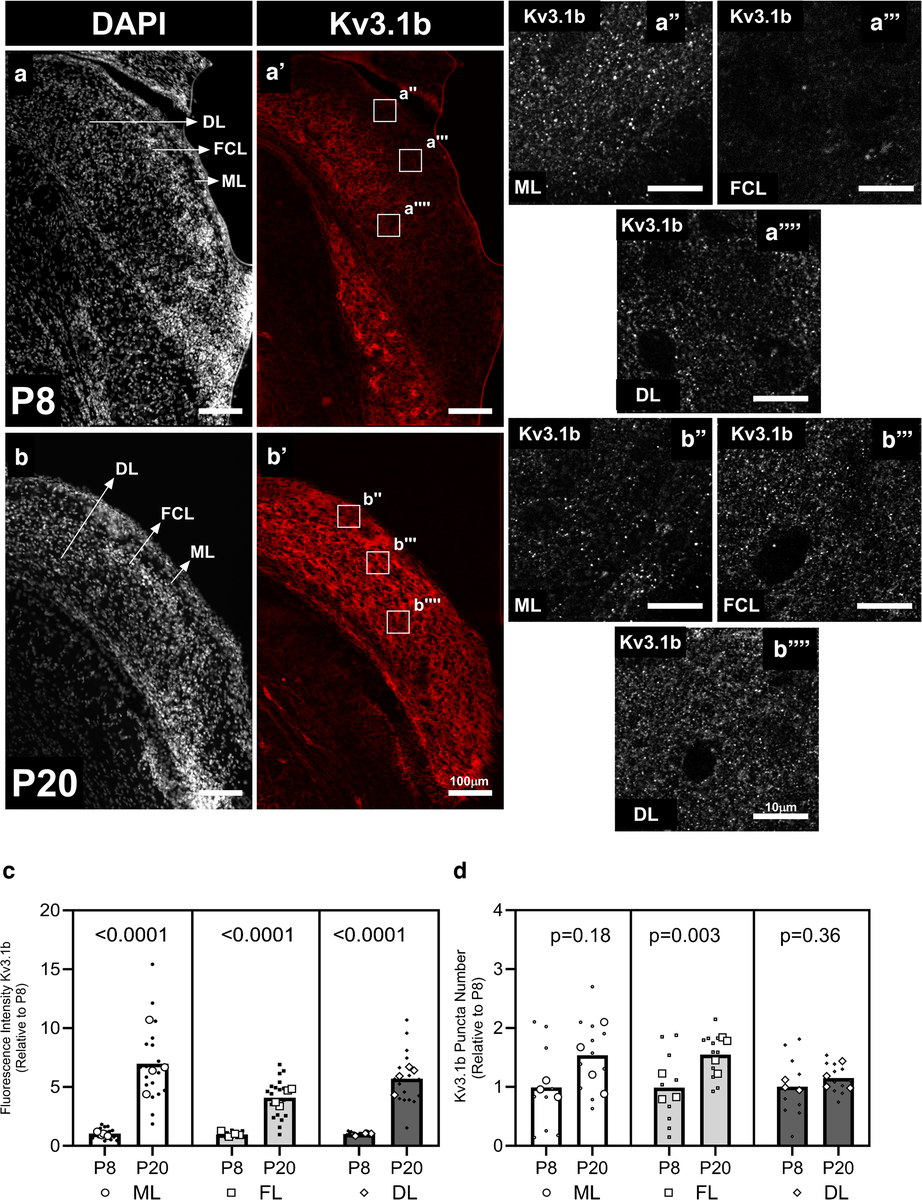
Figure 1. Kv3.1b protein (red) increases sharply across the dorsal cochlear nucleus after hearing onset (P20) compared to before (P8), especially in the fusiform cell layer.
However, when the team turned to voltage-clamp recordings to measure the actual currents flowing through these channels, the picture grew more complex. While total outward potassium currents were indeed larger in post-hearing neurons, the TEA-sensitive component,the pharmacological proxy for Kv3 current,was remarkably similar in magnitude between the two age groups. The significant increase was in the TEA-resistant potassium current, pointing to other channel families (potentially Kv1 or Kv2) as the main contributors to the larger total current seen after hearing onset.
Dissecting the Action Potential with Pharmacology
The most revealing experiments came from current-clamp recordings performed before and after application of 5 mM TEA to block Kv3 channels. If Kv3.1 channels were responsible for the developmental shortening of action potentials, then blocking them should eliminate the differences between pre- and post-hearing neurons. That is not what happened.
TEA broadened action potentials in both age groups,increasing amplitude, half-width, and decay time while decreasing the decay slope,confirming that Kv3 channels do contribute to repolarization at both ages. But critically, the developmental differences in action potential waveform persisted after TEA application. Post-hearing neurons still had faster, shorter spikes than pre-hearing neurons even when Kv3 channels were blocked. This elegant result demonstrates that Kv3.1 is not the channel responsible for the maturation of individual spike waveforms.
Where Kv3 channels did prove indispensable was in sustaining high-frequency firing. TEA reduced the maximum firing rate of post-hearing neurons significantly more than it affected pre-hearing neurons. Even more strikingly, TEA induced burst firing in 93% of post-hearing neurons compared to only 36% of pre-hearing neurons. This burst firing was not an artifact of blocking calcium-activated BK potassium channels,a possibility the team ruled out by showing that cadmium chloride blockade of calcium influx did not reproduce the bursting phenotype. These results paint a clear picture: Kv3.1b upregulation after hearing onset specifically equips fusiform neurons to fire at the sustained high rates required for processing temporal features of sound.
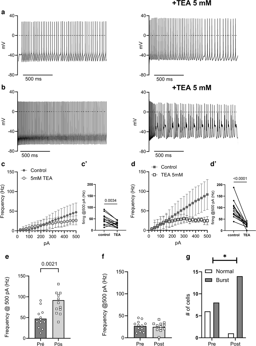
Figure 2. Blocking Kv3 channels with TEA disrupts normal, steady firing and triggers bursting activity in fusiform neurons after hearing onset, revealing Kv3’s role in sustaining high-frequency firing.
Precision Brainstem Slicing for Demanding Electrophysiology
The electrophysiological recordings in this study demanded exceptional slice quality. The researchers needed to obtain viable 200 µm coronal brainstem slices from postnatal mice as young as P8,tissue that is small, soft, and extraordinarily fragile. Fusiform neurons in the DCN sit within a layered structure that must remain intact for meaningful patch-clamp recordings, and any mechanical distortion during sectioning could compromise both cell health and the integrity of local circuitry.
To meet these demands, the team used a Compresstome VF-300-0Z vibrating microtome. The Compresstome‘s agarose embedding and compression tube system stabilizes tissue during sectioning without requiring adhesives or excessive manual handling. For delicate postnatal brainstem tissue, this approach is particularly valuable: the agarose provides uniform support around the specimen, minimizing the compression artifacts and cellular damage that can plague conventional vibratome preparations. The result is cleaner cut surfaces with healthier neurons at the slice face,a prerequisite for the whole-cell patch-clamp recordings that were central to every electrophysiological conclusion in this study.
The team also prepared 35 µm cryostat sections for their immunofluorescence work, but it was the Compresstome-prepared acute slices that enabled the functional experiments connecting Kv3.1b expression to its physiological consequences in living neurons.
Rewriting the Developmental Playbook for Auditory Brainstem Channels
This study challenges a straightforward narrative and replaces it with a more precise one. Kv3.1b channels are upregulated in DCN fusiform neurons after hearing onset, but their primary developmental contribution is enabling sustained high-frequency firing,not shaping the waveform of individual action potentials. The shortening and acceleration of spikes must be attributed to other potassium channel subtypes, with Kv1 and Kv2 families being strong candidates for future investigation.
The finding that TEA-induced burst firing disproportionately affects post-hearing neurons is particularly relevant for understanding tinnitus. If acoustic trauma reduces Kv3 currents in fusiform neurons,as prior work suggests,then the resulting shift toward burst firing could be a key mechanism driving the aberrant spontaneous activity associated with phantom sound perception. The work by de Oliveira and colleagues provides a refined framework for understanding exactly which aspect of neuronal function is disrupted when Kv3.1 channels are compromised.
We at Precisionary are proud that the Compresstome played a role in enabling this meticulous developmental neuroscience. If your research involves acute brain slice electrophysiology, auditory brainstem circuitry, or the study of ion channel contributions to neural development, we would love to hear about your work. Reach out to our team to learn how precision tissue sectioning can support your next discovery.