

Key Takeaways
- A new study from Beth Israel Deaconess Medical Center (BIDMC) and Harvard Medical School, generated with Precisionary’s Compresstome vibrating microtome, extends viable human precision-cut liver slice (PCLS) culture to 10 to 14 days, far beyond the field baseline where fewer than 7% of published PCLS studies maintain culture past 72 hours.
- During extended culture, healthy-donor slices developed bridging fibrosis by day 7, while cirrhotic-donor slices showed hepatocyte-to-biliary transdifferentiation (ductular reaction), recapitulating real human liver disease biology.
- Precisionary’s Chief Scientific Officer, Abby Chu, is a co-author on the paper.
A new research letter published in eGastroenterology, “Extended precision cut liver slice culture models liver regeneration and ductular reaction,” reports one of the longest viable human liver slice cultures described to date. The study, led by Sofia Izunza Barba and colleagues, with senior author Z. Gordon Jiang, MD, PhD (Division of Gastroenterology, BIDMC and Harvard Medical School), used the Compresstome vibrating microtome to generate precision-cut liver slices (PCLS) from 25 human explant livers, iterating the protocol over two years to push culture viability well past the field’s usual limits.

Supplementary Figure 1. Tissue embedding and sectioning on the Compresstome. A: liver tissue cored and embedded in agarose. B: the agarose-embedded tissue sectioned into 250 µm slices on the Compresstome VF-510-0Z. C: uniform PCLS transferred to culture. From Izunza Barba S, et al. eGastroenterology 2026;4:e100389. CC BY-NC 4.0.
Research Question and Background
Precision-cut liver slice technology has existed for more than four decades, but adoption has been limited by short culture lifespans, restricted access to viable human tissue, and reproducibility concerns. A 2021 review found that only 30% of PCLS studies used human liver tissue at all, and fewer than 7% maintained cultures beyond 72 hours. The Jiang lab set out to build a more durable, better-validated human PCLS platform using a tissue source most labs overlook: explant livers removed during transplantation, spanning donor biology from MASH, alcohol-associated hepatitis, ALD, HBV, and PSC to non-fibrotic healthy controls.
- Establish a refined PCLS protocol using explant human liver tissue rather than scarce healthy donor tissue
- Extend viable culture duration well beyond the field’s 72-hour norm
- Develop non-destructive, real-time viability benchmarks to guide protocol optimization
- Characterize how the tissue remodels over extended culture, in both healthy and cirrhotic donor liver
Key Findings
Using three complementary viability benchmarks, whole-plate morphology imaging, whole-mount dual-color live/dead staining, and Seahorse oxygen consumption analysis, the team verified viable PCLS culture out to 10 to 14 days. Along the way, they found that healthy and cirrhotic donor tissue behave very differently in extended culture:
- Healthy-donor PCLS developed portal fibrosis progressing to bridging fibrosis by day 7, and formed regenerative hepatocyte colonies lacking basement membrane after day 7.
- Cirrhotic-donor PCLS instead showed a gradual emergence of KRT19-positive biliary/progenitor cells adjacent to myofibroblasts, a ductular reaction that mirrors hepatocyte-to-biliary transdifferentiation seen in cirrhosis and alcohol-associated hepatitis; this regeneration pattern was not seen in the cirrhotic tissue.
- Oxygen consumption was higher in healthy-donor slices than cirrhotic-donor slices at day 3, then diverged further over time: declining in healthy tissue but rising in cirrhotic tissue, pointing to distinct patterns of cellular remodeling between the two.

Supplementary Figure 3. Whole-mount live (green, Calcein AM) / dead (red, EthD-III) viability imaging of PCLS from healthy and cirrhotic donor liver across the culture time course. From Izunza Barba S, et al. eGastroenterology 2026;4:e100389. CC BY-NC 4.0.
The team also demonstrated that explant livers, often cirrhotic, but sometimes from polycystic liver disease or metastatic cancer in remission, are a practical and underused source of both diseased and near-normal “healthy” control tissue.
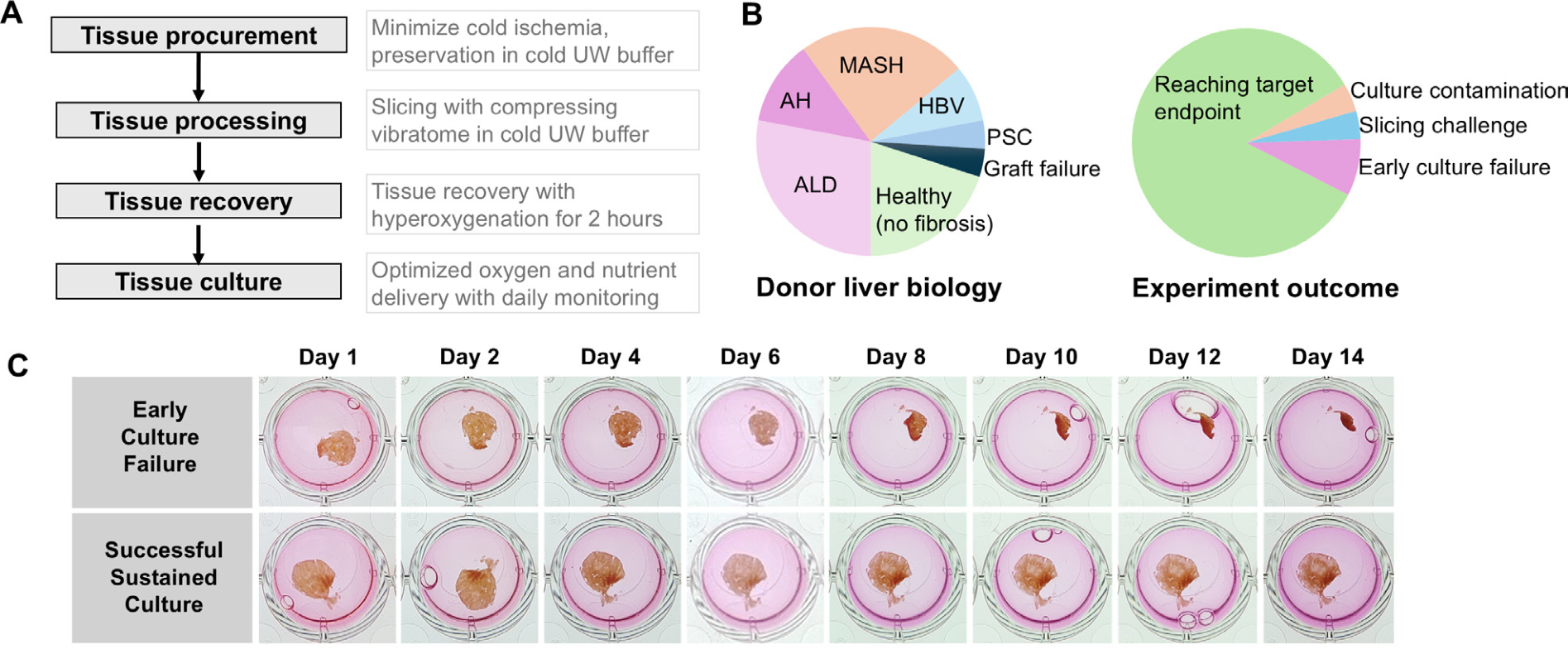
Figure 1 (panels A–C). A: the refined PCLS generation workflow. B: donor liver biology and experiment outcomes across the 25 processed livers. C: whole-plate imaging comparing early culture failure to successful sustained culture over 14 days. From Izunza Barba S, et al. eGastroenterology 2026;4:e100389. CC BY-NC 4.0.
How the Compresstome Supported This Research
Fresh liver tissue was cored, embedded in agarose, and sectioned on a Compresstome VF-510-0Z vibrating microtome under chilled University of Wisconsin (UW) preservation buffer, producing uniform 250 µm slices. The authors specifically credited the compression-assisted slicing approach with producing more uniform sections than earlier PCLS methods, a key input to their extended-viability result.
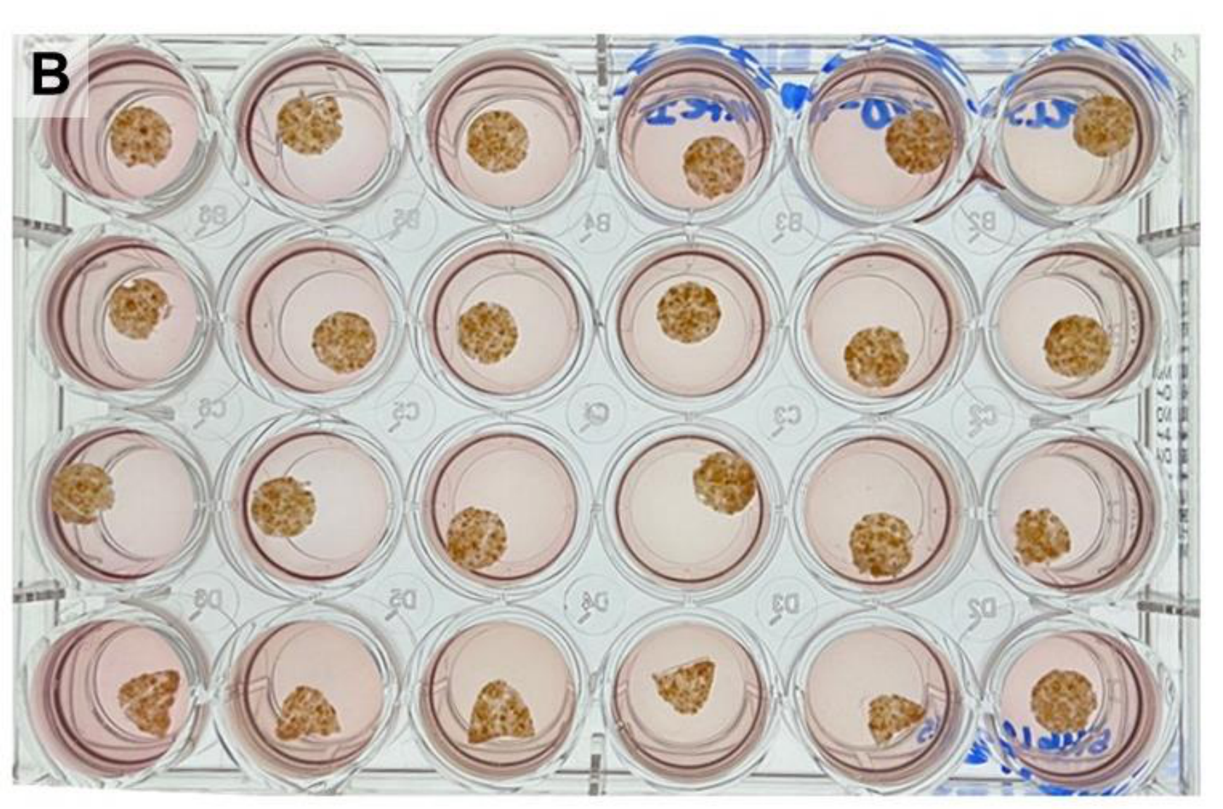
What you can get with PCLS using the Compresstome: a full 24-well plate of intact human liver slices, freshly sectioned and ready for extended culture. Adapted from Supplementary Figure 2B, Izunza Barba S, et al. eGastroenterology 2026;4:e100389. CC BY-NC 4.0.
Why It Matters
Precision-cut liver slices are a canonical New Approach Methodology (NAM) under the NIH’s push to reduce reliance on animal models while generating more human-relevant data. A two-week viability window changes what’s practically possible with PCLS, opening the door to longer-duration studies, including repeat-dose and fibrosis-progression work, in intact human tissue architecture rather than a single 72-hour snapshot.
We’re proud to support research like this. If your work touches liver toxicology, fibrosis, or disease modeling, we would love to hear what you’re working on.
→ Read the full paper
→ Request our liver PCLS protocol
→ Talk to our team