

Key Takeaways
- The tail striatum (TS) is a poorly understood brain region linked to sensory processing, and its medium spiny neurons (MSNs) have never been characterized electrophysiologically in depth — until now.
- TS-MSNs are electrophysiologically distinct from dorsolateral striatum (DLS) MSNs, with more depolarized resting potentials, lower rheobase, and stronger spontaneous excitatory input.
- The three anatomical divisions of the tail striatum — medial, intermediate, and lateral — also differ functionally and respond unevenly to dopamine and selective dopamine receptor agonists.
- These findings have direct implications for understanding how dopamine dysfunction in the tail striatum may drive sensory symptoms such as hallucinations in Parkinson’s disease and schizophrenia.
- The researchers used the Compresstome vibrating microtome to prepare 200 µm acute brain slices from juvenile rats, enabling the precise whole-cell patch clamp recordings that made this first-of-its-kind characterization possible.

Fig. 2a from Riley et al. (2026), Experimental Physiology. Medium spiny neuron somas imaged live in Compresstome-prepared acute tail striatum slices, immediately before whole-cell patch-clamp recording.
Most neuroscience research on the striatum has focused on its rostral and dorsal regions — areas long known to be critical for motor control and reward. But the tail end of the striatum, tucked into the caudal brain, has remained something of a terra incognita. In a new paper published in Experimental Physiology, Riley et al. (2026) deliver the first comprehensive electrophysiological portrait of medium spiny neurons across the tail striatum’s three anatomical divisions, revealing a functionally diverse landscape with distinct dopaminergic modulation — and opening a new window into sensory symptoms of diseases like Parkinson’s and schizophrenia.
Why the Tail Striatum Has Been a Blind Spot
The striatum is the primary input nucleus of the basal ganglia, and its role in generating behavior and shaping perception is well established. The majority of its neurons — roughly 95% — are medium spiny neurons (MSNs), which are broadly classified by their expression of D1 or D2 dopamine receptors. In the rostral striatum, D1- and D2-MSNs are intermingled and their distinct electrophysiological and pharmacological profiles have been studied extensively. These properties are foundational to models of how dopamine dysfunction leads to motor symptoms in Parkinson’s disease and to reward-processing abnormalities in addiction.
The tail striatum (TS), however, tells a different organizational story. Rather than intermingling D1- and D2-expressing neurons, the TS is anatomically segregated into medial, intermediate, and lateral divisions with uneven dopamine receptor expression: the medial division is D1-rich, the intermediate division is D2-rich, and the lateral division expresses both. This unusual arrangement has been documented anatomically, but what it means for neural function has been a mystery. No one had performed a detailed electrophysiological characterization of TS-MSNs, let alone tested how dopamine and selective receptor agonists modulate these neurons across the three divisions. Riley and colleagues set out to fill that gap.
A First-of-Its-Kind Electrophysiological Map
Working with brain slices from juvenile Wistar rats (postnatal days 45–55, both sexes), the research team used whole-cell patch clamp electrophysiology to systematically record from MSNs in each TS division and in the adjacent dorsolateral striatum (DLS), which served as a well-characterized comparison region. They also performed immunofluorescence staining to confirm D1 and D2 receptor expression patterns and used pharmacological experiments to probe dopaminergic modulation.
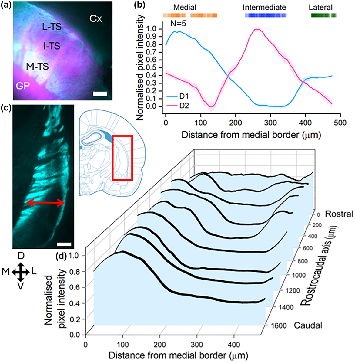
Fig. 1 from Riley et al. Mapping D1 and D2 dopamine receptor ‘stripes’ across the tail striatum reveals its hidden internal structure.
The results revealed that TS-MSNs are fundamentally different from their DLS counterparts. Across a suite of electrophysiological measures, TS-MSNs showed more depolarized resting membrane potentials, lower membrane capacitance despite having larger and rounder somas, lower rheobase (meaning they required less current to fire), and higher frequency and amplitude of spontaneous excitatory postsynaptic currents (sEPSCs). Taken together, these properties paint a picture of neurons that sit closer to firing threshold and receive stronger excitatory drive — consistent with a region that may need to respond rapidly to sensory input.
But the story didn’t end at the TS-vs-DLS level. Within the tail striatum itself, the three divisions showed meaningful functional heterogeneity. The intermediate division had a higher rheobase than the medial or lateral divisions, suggesting it is less easily excited. The lateral division stood out for higher firing frequencies at rheobase compared to both the medial and intermediate divisions, and for larger action potential amplitudes compared to the medial division specifically. When the team applied Fisher’s linear discriminant analysis to the electrophysiological data, the classifier could successfully distinguish medial and lateral TS-MSNs from neurons in other divisions — but struggled with the intermediate division, suggesting greater intrinsic heterogeneity there, perhaps reflecting its transitional position between the D1-rich medial and mixed lateral zones.
Dopamine Doesn’t Act the Same Way Everywhere
One of the most striking findings concerned how dopamine modulates MSN activity across these regions. When the researchers bath-applied dopamine at 30 and 100 µM concentrations, it suppressed firing in a subset of MSNs — but this suppression was far from uniform. DLS-MSNs were the most commonly affected, followed by medial TS-MSNs. Lateral division MSNs were rarely suppressed, and intermediate division MSNs were not suppressed at all.
Selective agonist experiments sharpened the picture further. The D1 agonist SKF38393 modulated firing only in the D1-rich medial division, while the D2 agonist quinpirole affected only the D2-rich intermediate division. In both cases, the modulation was weaker than what is typically observed in the DLS. This division-specific pharmacology aligned beautifully with the known anatomical segregation of dopamine receptor subtypes — but it also revealed something unexpected. Despite their segregated receptor expression, the D1-rich medial and D2-rich intermediate TS divisions shared similar resting membrane properties — a marked contrast to the rostral striatum, where D1- and D2-MSNs differ substantially on this front despite being intermingled. (The two divisions still differ in firing properties, such as rheobase.) This suggests that the organizational logic of the tail striatum may differ fundamentally from that of its rostral counterpart.
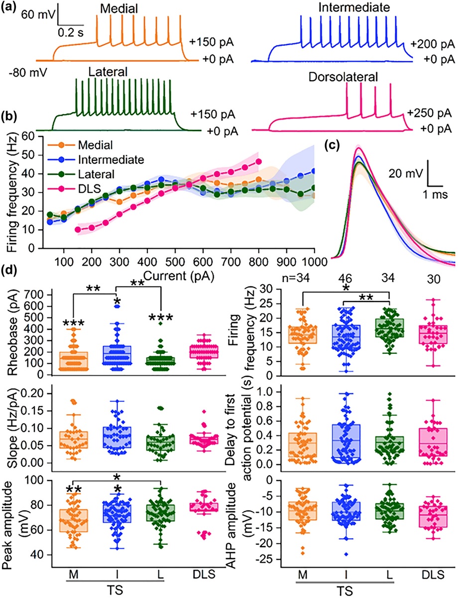
Fig. 4 from Riley et al. Neurons in different striatal regions fire differently: recordings show distinct firing patterns and action potential shapes across striatal divisions.
Precision Sectioning for Precision Electrophysiology
This kind of work — recording from identified neurons within specific sub-regions of a small, caudally located brain structure — demands exceptional slice quality. The tail striatum is compact, and distinguishing its medial, intermediate, and lateral divisions requires slices that preserve both the cytoarchitecture needed for anatomical identification and the cellular health needed for stable whole-cell recordings. Compression artifacts, uneven surfaces, or damaged superficial cell layers can make the difference between a successful recording session and a wasted preparation.
Riley et al. prepared their 200 µm coronal brain slices using the Compresstome VF-300-02 vibrating microtome. The Compresstome‘s agarose compression tube system stabilizes tissue during sectioning by surrounding the specimen in a cylinder of solidified agarose, which provides uniform support on all sides. This approach minimizes the lateral displacement and compression artifacts that conventional vibratomes can introduce — artifacts that are particularly problematic for electrophysiology, where even subtle mechanical damage to superficial neurons can reduce the yield of viable patch clamp recordings. For a study that needed to record from dozens of MSNs across four distinct sub-regions, each requiring precise anatomical targeting, consistent slice quality wasn’t a luxury — it was a prerequisite.
From Electrophysiology to Understanding Hallucinations
Beyond its contribution to fundamental striatal neuroscience, this work carries real translational weight. Sensory symptoms — including visual and auditory hallucinations — are common in both Parkinson’s disease and schizophrenia, yet they remain poorly understood at the circuit level. The tail striatum, with its predominantly sensory inputs and its dependence on dopaminergic modulation, is a prime candidate for mediating these symptoms. By showing that TS-MSNs respond differently to dopamine than DLS-MSNs, and that even within the TS the three divisions have distinct pharmacological profiles, Riley et al. provide a mechanistic framework for understanding how localized dopamine dysfunction could selectively disrupt sensory processing without necessarily affecting motor function.
The intermediate division — the most D2-rich — offers a particularly intriguing case. None of its neurons were fully silenced by dopamine, unlike a subset of neurons in the other regions. Yet among neurons that kept firing, the intermediate division actually showed the strongest dopaminergic modulation of any TS division: firing frequency dropped significantly at both dopamine concentrations tested, and rheobase rose significantly at the higher dose. In other words, dopamine doesn’t shut these neurons down — it fine-tunes how readily they fire. This suggests that dopaminergic modulation of this division operates through more nuanced mechanisms than simple on/off silencing, with potential implications for how antipsychotic drugs (most of which target D2 receptors) affect sensory circuits.
This study is a foundational contribution to an emerging field. As researchers continue to dissect the functional architecture of the tail striatum, the electrophysiological and pharmacological baselines established here will serve as essential reference points. We’re proud that the Compresstome played a role in enabling this meticulous work.
If your research involves acute brain slice electrophysiology, striatal circuitry, or dopaminergic modulation — particularly in understudied regions like the tail striatum — we’d love to hear about your work. Reach out to our team to discuss how precision tissue sectioning can support your next project.