

A new study from Yale University, published in Science Advances (Ziolkowski et al.), overturns a decades-old model of how the Pacinian corpuscle, the receptor responsible for detecting transient touch and high-frequency vibration in skin, actually works. Led by Elena Gracheva and Sviatoslav Bagriantsev, the team used the Compresstome to prepare tissue for high-resolution electron microscopy, revealing that the corpuscle’s “inner core” does far more than support its sensory nerve terminal: its cells are active, mechanosensitive contributors to touch detection in their own right.
Key Takeaways
- Using eFIB-SEM imaging of Compresstome-sectioned duck bill skin, researchers built the first nanoscale (8-nm resolution) 3D reconstruction of a Pacinian corpuscle, the touch/vibration receptor found in skin.
- The study overturns the long-held model that the corpuscle’s outer capsule alone tunes its sensitivity to touch and vibration. That filtering role turns out to be dispensable.
- Lamellar Schwann cells (LSCs) inside the corpuscle’s inner core are themselves mechanosensitive, and activating them lowers the touch-detection threshold of the sensory nerve terminal they surround.
- The Compresstome cleanly sectioned fixed embryonic duck bill skin into uniform 300-µm slices, the critical first step that made high-resolution volumetric electron microscopy of an intact corpuscle possible.
Research Question / Background
Pacinian corpuscles are layered structures wrapping a sensory nerve terminal, tuned specifically to detect vibration and transient touch rather than sustained pressure. For decades, the accepted model held that a multilayered “outer core” acts as a mechanical filter, damping static and low-frequency forces so only fast, transient stimuli reach the terminal, while the “inner core” (lamellar Schwann cells, or LSCs, wrapping the terminal) was assumed to play a purely structural role. But prior work had shown the corpuscle’s tuning persists regardless of how many outer-core layers remain, raising doubt about that model. This study set out to:
- Reconstruct the full 3D ultrastructure of a Pacinian corpuscle at nanometer resolution, including the outer core, the inner-core LSCs, and the afferent nerve terminal
- Test whether disrupting the outer core changes the corpuscle’s signature rapid adaptation and high-frequency filtering
- Determine, via direct patch-clamp recording, whether LSCs are themselves mechanosensitive
- Establish whether LSC activity is functionally coupled to the nerve terminal’s touch threshold
Key Findings
- Built the first 8-nm-resolution 3D reconstruction of an intact Pacinian corpuscle, resolving the outer core’s lamellar cells, 12 individual inner-core LSCs, and the full architecture of the nerve terminal they surround.
- Disrupting the outer core did not eliminate rapid adaptation or high-pass frequency filtering. The corpuscle’s tuning persisted, challenging the accepted “mechanical filter” model.
- Patch-clamp recordings showed LSCs generate their own mechanically activated currents, direct evidence that these cells are mechanosensitive, not merely structural.
- Activating LSCs lowered the mechanical threshold needed to trigger a response in the nerve terminal, and electron microscopy revealed extensive physical contacts (junctions and tethers) between LSC lamellae and the terminal membrane, evidence of tight functional coupling between the two cell types.
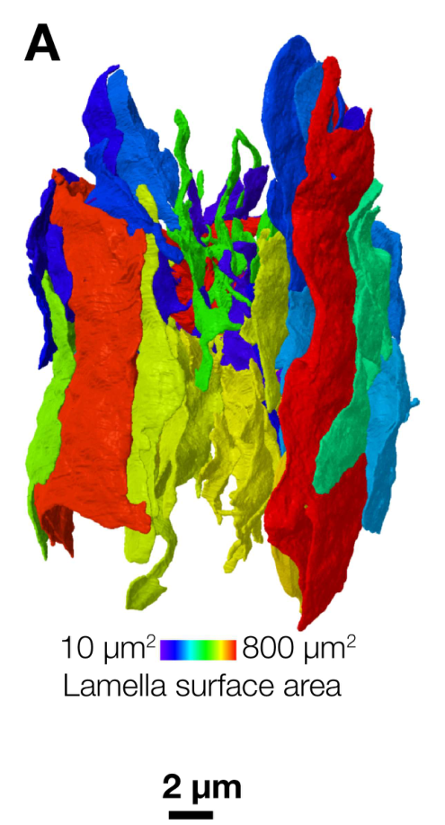
Figure S2. A single lamellar Schwann cell (LSC) and its lamellae, color-coded by surface area. Ziolkowski et al., Science Advances (2025).
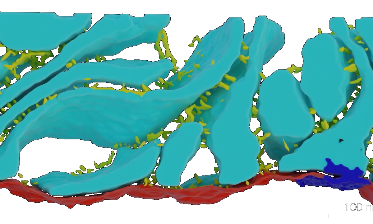
Movie S2 (still). Junctions and tethers (yellow) connecting LSC lamellae (cyan) to the afferent terminal (red). Ziolkowski et al., Science Advances (2025).
How the Compresstome Enabled This Research
To reach nanometer-scale imaging, the team first needed intact, uniformly thin tissue sections free of the compression or tearing artifacts that can distort delicate corpuscle ultrastructure. They used the Compresstome to cut fixed embryonic Mallard duck bill skin into 300-µm sections, which were then stained, resin-embedded, and imaged by enhanced focused ion beam scanning electron microscopy (eFIB-SEM) at 8-nm³ resolution. That clean, reproducible sectioning was the necessary starting point for the paper’s flagship reconstruction: without uniform, undamaged slices at that thickness, the downstream nanoscale imaging pipeline wouldn’t have been possible.
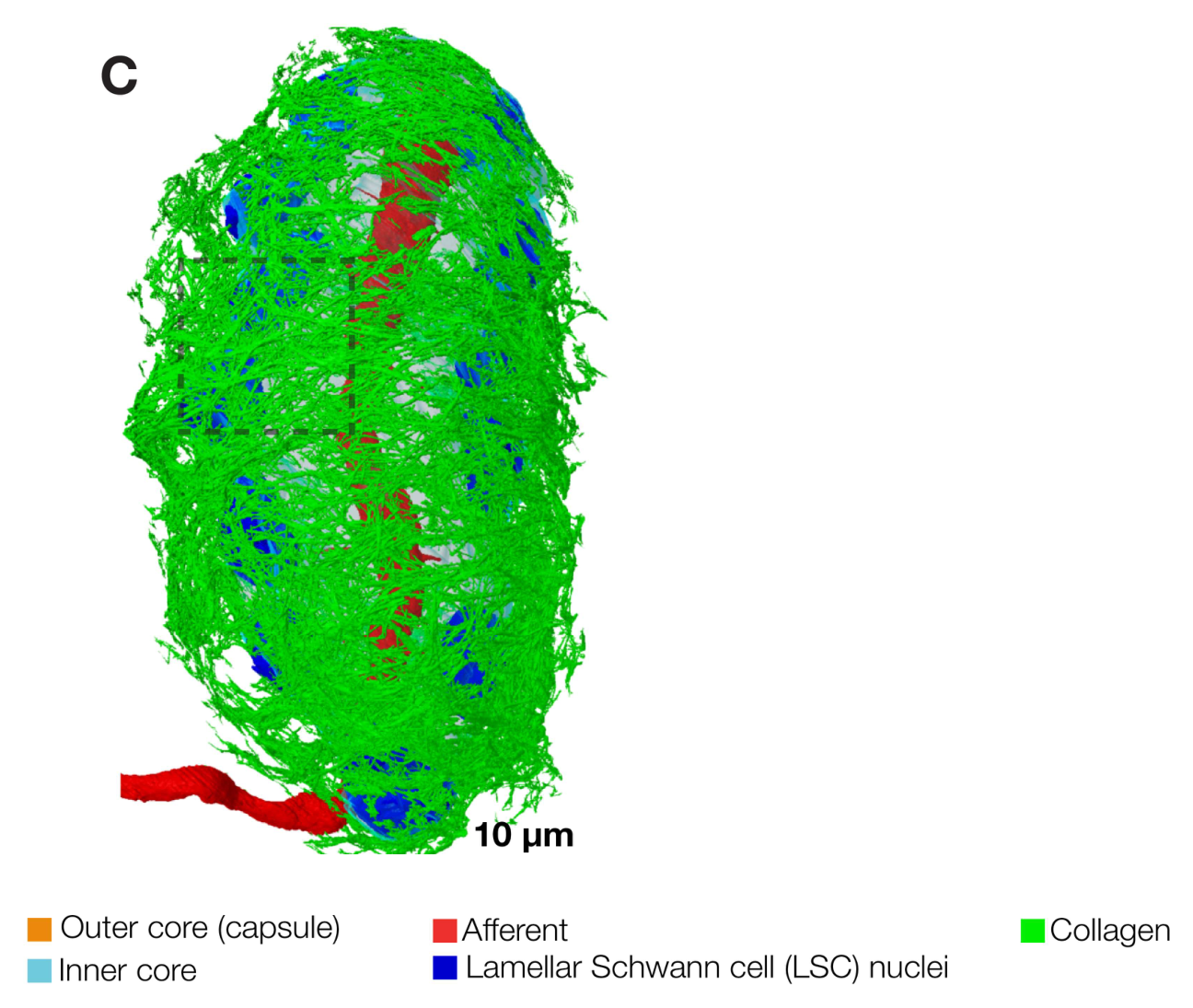
Figure S1. 3D reconstruction of a Pacinian corpuscle from Compresstome-sectioned duck bill skin, imaged by enhanced FIB-SEM. Outer core (orange), inner core (cyan), afferent terminal (red), LSC nuclei (blue), collagen (green). Ziolkowski et al., Science Advances (2025).
Why It Matters
This work reframes Schwann cells inside the Pacinian corpuscle from passive scaffolding into active sensory participants, a shift that could reshape how researchers think about mechanotransduction more broadly, across touch, proprioception, and vibration sensing. Because Pacinian corpuscles are large, individually identifiable, and accessible in duck bill skin, this system offers a rare chance to directly pair structural and functional study of a single sensory receptor, a combination that’s difficult in most other touch-sensing tissue.
We’re proud to see the Compresstome supporting foundational neuroscience research like this, helping preserve the delicate tissue architecture needed to answer fundamental questions about how we sense the world around us.