

Key Takeaways
- Two doses of psilocybin administered before chemotherapy prevented peripheral neuropathy in mice for up to eight months, without impairing the anti-tumor efficacy of the chemotherapy itself.
- The neuroprotective effect required serotonin 2A receptor activation but not the drug’s hallucinogenic properties. A nonhallucinogenic 2A agonist worked equally well, opening a practical path to clinical translation.
- Psilocybin preserved axonal mitochondrial transport through a TrkB, Akt, PAK5, MAP2, KIF5B signaling cascade, a mechanism confirmed in live human peripheral nerve tissue and human stem cell-derived sensory neurons.
- Acute medial prefrontal cortex (mPFC) brain slices prepared with the Compresstome vibrating microtome enabled multielectrode array recordings that revealed how chemotherapy disrupts cortical network activity and how psilocybin restores it.
- With no current FDA-approved preventive treatment for chemotherapy-induced peripheral neuropathy, these findings position psilocybin and related compounds as a first-in-class prophylactic strategy ready for rigorous clinical evaluation.
Chemotherapy-induced peripheral neuropathy (CIPN) affects a staggering proportion of cancer patients receiving platinum- and taxane-based regimens. It causes numbness, burning pain, and loss of fine motor function in the hands and feet, symptoms that often persist long after treatment ends and for which no preventive therapy exists. In a study published in Science, Heles et al. (2026) report that psilocybin, administered prophylactically before chemotherapy, prevents neuropathy from developing in mouse models and trace the mechanism to the preservation of mitochondrial transport along peripheral sensory axons, a finding they validate in living human nerve tissue. The scope of the work, spanning behavioral pharmacology, electrophysiology, live-cell imaging, molecular biology, and human translational experiments, makes it one of the most comprehensive mechanistic studies of neuroprotection in recent memory.
An Unmet Clinical Need Hiding in Plain Sight
Despite decades of research, CIPN remains one of the most common and undertreated toxicities of cancer therapy. Platinum drugs such as cisplatin damage the sensory neurons of the dorsal root ganglia (DRG), while taxanes like paclitaxel disrupt microtubule dynamics essential for axonal transport. In both cases, the result is the same: dying-back degeneration of distal nerve endings, energy failure at the periphery, and chronic neuropathic pain that can force dose reductions or treatment discontinuation. The lack of a preventive strategy is not for lack of trying: trial after trial of putative neuroprotective agents has failed, in part because the field has lacked a clear mechanistic target. Heles and colleagues set out to change that by testing whether psilocybin, a serotonin 2A receptor agonist with an established clinical safety profile, could intervene upstream of neuropathy onset and by defining exactly how it does so.
Prophylactic Protection That Lasts Across Chemotherapy Cycles
The central behavioral finding of the study is striking in its durability. In multiple CIPN mouse models, using cisplatin as well as the taxanes paclitaxel and docetaxel, just two doses of psilocybin given before the first chemotherapy cycle prevented the development of mechanical pain hypersensitivity. Crucially, this protection was not transient: it held across six consecutive chemotherapy cycles and persisted for eight months of follow-up. The authors also demonstrated that psilocybin did not interfere with the chemotherapy’s anti-tumor activity, nor did it alter systemic immune cytokine profiles in tumor-bearing mice. This last point is essential for clinical viability, since any neuroprotective strategy must not compromise the cancer treatment it accompanies.
To dissect whether psilocybin’s well-known psychoactive properties are part of the neuroprotective mechanism, the team tested a nonhallucinogenic serotonin 2A receptor agonist and found it equally effective. They then asked where in the nervous system the receptor activation needed to happen. Blocking serotonin 2A receptors systemically abolished psilocybin’s protection of tactile function, but blocking them centrally, via direct intracerebroventricular injection into the brain, did not. That distinction points to peripheral serotonin 2A signaling, not central, as the key driver of psilocybin’s protection against tactile deficits. Together, these findings indicate that the protective pharmacology can potentially be delivered without the psychoactive experience, and that it works through the peripheral nervous system rather than requiring a central, brain-wide effect, which could meaningfully lower the barriers to prophylactic use in oncology clinics.
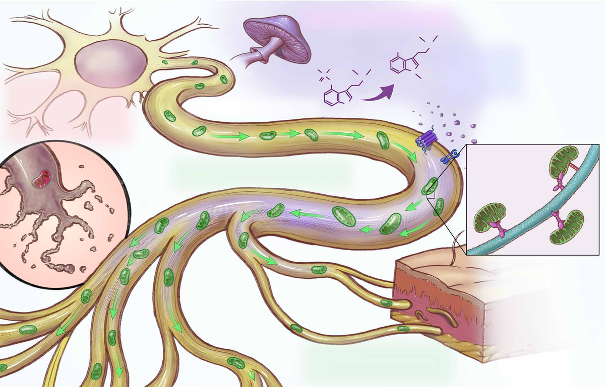
Figure 1. Psilocybin helps keep mitochondria moving along nerve fibers, powering the nerve endings that chemotherapy typically damages. Source: Heles et al., Science (2026).
Tracing the Mechanism from Cortex to Axon Terminal
What makes this paper exceptional is how thoroughly it maps the mechanism at both the central and peripheral levels. Centrally, the authors used electroencephalography (EEG) in awake mice to show that cisplatin disrupts cortical oscillatory patterns, a finding consistent with the “chemobrain” cognitive effects reported by patients. They then turned to acute brain slice electrophysiology for a more granular view of cortical network function. Using 250-micron acute slices of the medial prefrontal cortex (mPFC) prepared with the Compresstome vibrating microtome, they performed multielectrode array (MEA) recordings that revealed a significant reduction in network firing rate in cisplatin-treated animals. Psilocybin pretreatment restored this activity toward vehicle control levels, corroborating the in vivo EEG normalization and providing direct evidence that psilocybin preserves cortical circuit function against chemotherapy-induced disruption.
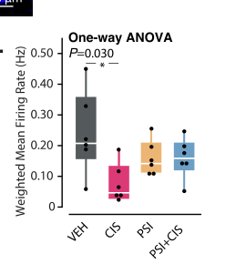
Weighted mean firing rate from multielectrode array recordings of Compresstome-prepared mPFC slices. Cisplatin (CIS) reduces network firing rate relative to vehicle (VEH); psilocybin pretreatment (PSI+CIS) restores activity toward vehicle levels. Source: Heles et al., Science (2026), Fig. 2L.
Peripherally, the story converges on mitochondrial trafficking. Using live-cell imaging of DRG neurons and, remarkably, freshly isolated living human peripheral nerve tissue, the researchers showed that chemotherapy stalls the active transport of mitochondria along sensory axons. Without a steady supply of mitochondria reaching the distal nerve terminals, those endings lose their energy supply and degenerate. Psilocybin prevented this trafficking deficit. The molecular cascade responsible, TrkB activation leading through Akt, PAK5, MAP2, and the kinesin motor KIF5B, was confirmed using pharmacological inhibitors (including the TrkB inhibitor ANA-12) and validated in human iPSC-derived sensory neurons and patient skin biopsies from the TRIP clinical trial cohort. This is not a single-experiment mechanism claim; it is a signaling pathway triangulated across species and experimental systems.
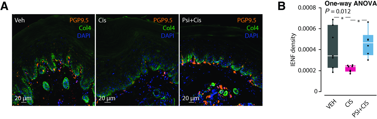
Figure 2. Skin nerve fibers (orange) are depleted by chemotherapy alone, but preserved when psilocybin is given beforehand. Source: Heles et al., Science (2026).
How Precision Brain Sectioning Supported the Electrophysiology
The acute mPFC slice experiments were a linchpin of the study’s central mechanism story, and the quality of those recordings depended directly on the quality of the tissue sections. Preparing viable 250-micron acute brain slices for multielectrode array work demands consistent thickness, minimal tissue compression, and intact cytoarchitecture. Any deformation of the cortical layers can distort network firing patterns and introduce artifacts into the very measurements the experiment is designed to capture. The researchers used the Compresstome VF-510-0Z for this preparation. The instrument stabilizes the tissue block in an agarose embedding within a compression tube during sectioning, supporting the kind of uniform, low-artifact slices that multielectrode array work depends on, where electrode-tissue contact across the entire array must be consistent and the slice must remain viable over extended recording sessions. The clarity of the network-level firing rate differences the authors report between cisplatin, psilocybin-pretreated, and vehicle groups speaks to the integrity of the slice preparations underlying those measurements.
From Bench to Bedside: What This Means for Cancer Patients
The translational implications of this work are hard to overstate. CIPN currently has no FDA-approved prevention. Duloxetine, the only agent with even modest evidence for treating established CIPN, does not prevent it and offers limited relief. Psilocybin, meanwhile, already has a well-characterized safety profile from clinical trials in depression, end-of-life anxiety, and substance use disorders. The demonstration that a nonhallucinogenic serotonin 2A agonist provides equivalent protection removes perhaps the largest practical obstacle to prophylactic use, since it means neuroprotection could be achieved with a simple oral medication that does not require supervised psychedelic sessions. The identification of a defined molecular pathway, from TrkB through KIF5B-mediated mitochondrial transport, also opens the door to biomarker-guided patient selection and to the development of entirely new drug classes targeting axonal energy logistics.
Perhaps most compelling is the validation in human tissue. By showing that the mitochondrial trafficking mechanism is conserved in live human peripheral nerves, human stem cell-derived sensory neurons, and patient biopsy material, Heles and colleagues have built a bridge between the animal pharmacology and the human disease that is far sturdier than most preclinical neuroprotection studies manage to construct. Clinical trials evaluating prophylactic psilocybin or nonhallucinogenic 2A agonists in patients beginning neurotoxic chemotherapy regimens now have a clearly defined mechanistic rationale, validated endpoints, and a realistic path forward.
We at Precisionary Instruments are proud that the Compresstome played a role in enabling the electrophysiological experiments that helped define the central mechanism of this study. If your research involves acute brain slice electrophysiology, peripheral nerve studies, or precision tissue sectioning for neuroscience and translational oncology, we would love to hear about your work. Reach out to our team to discuss how the Compresstome can support your next study.