

Key Takeaways
- Researchers profiled how SMARCB1-deficient sinonasal carcinoma responds to drugs directly in patient tumor tissue, preserving the spatial architecture rather than dissociating the sample.
- Live tumor slices were generated on a Compresstome tissue slicer, producing uniform, viable sections suitable for ex vivo drug exposure.
- Drug responses varied across distinct regions of the same tumor, showing why spatially resolved profiling captures signals that bulk assays miss.
- The approach points toward functional, patient-specific testing for a cancer subtype with few effective treatment options.
Rare cancers present some of the most frustrating challenges in oncology — not because they are inherently less treatable, but because there are so few cases, so few models, and so little data to guide therapeutic decisions. SMARCB1-deficient sinonasal carcinoma (SDSC) is a textbook example: a highly aggressive malignancy of the nasal cavity and paranasal sinuses with dismal outcomes and virtually no preclinical platform to test potential therapies. A landmark new study published in EMBO Molecular Medicine by Jurmeister et al. (2026) takes a radically integrative approach to this problem, combining single-nucleus RNA sequencing, spatial transcriptomics, and patient-derived ex vivo tissue slice cultures to dissect the biology of SDSC at unprecedented resolution — and to identify actionable treatment strategies in living tumor tissue sliced on a Compresstome® VF-310-0Z vibrating microtome.
The result is the most comprehensive molecular and functional characterization of SDSC to date, and a compelling proof of concept for how precision tissue sectioning can power drug discovery in rare cancers that lack conventional cell line or animal models.
Why SDSC Has Been a Black Box — and Why That Needed to Change
SMARCB1 (also known as INI1) is a core subunit of the SWI/SNF chromatin remodeling complex, and its loss drives a small family of notoriously aggressive tumors, including malignant rhabdoid tumors in children and epithelioid sarcomas. SDSC was only formally recognized as a distinct entity in the last decade, and its rarity — with only scattered case series in the literature — has meant that basic questions about its cellular composition, spatial organization, and treatment vulnerabilities have remained unanswered.
There are no established cell lines for SDSC. There are no patient-derived xenograft models. Clinicians treating SDSC have essentially been working without a map, borrowing regimens from other head and neck cancers or SWI/SNF-mutant tumors with no direct evidence of efficacy. Jurmeister and colleagues set out to change this by building a comprehensive, multimodal atlas of an index SDSC tumor and then using that atlas to interpret the results of direct drug testing on living tissue slices.
Mapping Four Malignant States and Their Spatial Niches
Using single-nucleus RNA sequencing, the team resolved the tumor’s malignant compartment into four transcriptionally distinct populations: a predominant main cluster, a cycling subpopulation, and two specialized states marked by expression of ALDH1A1 and NTN4, respectively. This wasn’t just an exercise in cataloging — spatial transcriptomics (via the 10x Genomics Xenium platform) and sequential immunofluorescence revealed that these populations are not randomly distributed but instead occupy defined tissue niches.
Six spatially distinct niches were identified across the tumor. The ALDH1A1+ cells were particularly intriguing: they localized to a basal, stroma-proximal niche where they were intermingled with non-neoplastic p63-positive basal cells. Transcriptional analysis revealed that this ALDH1A1+ compartment displayed stem-like features with reduced proliferative activity — a profile that often signals therapeutic resistance in other tumor types, but which in this case harbored an unexpected vulnerability.
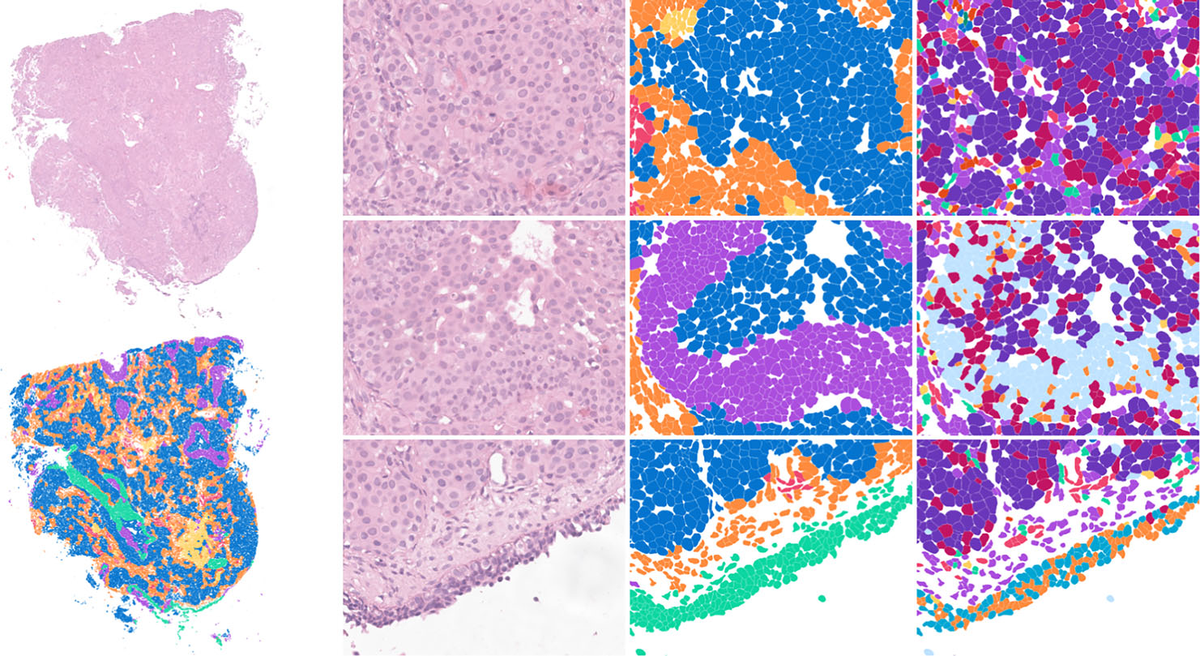
Figure 1. Mapping tumor tissue at single-cell resolution: spatial transcriptomics reveals distinct cellular neighborhoods within sinonasal carcinoma, distinguishing tumor cells from surrounding basal and stromal populations.
Sapanisertib Strikes at the Heart of Tumor Heterogeneity
The study’s most clinically consequential results came from the ex vivo tissue slice culture (TSC) drug testing platform. Fresh tumor tissue was sectioned into 300 µm slices using the Compresstome® and cultured under controlled conditions to preserve the native tissue architecture, microenvironment, and cellular diversity of the living tumor. These slices were then treated with a panel of candidate therapeutics, including the mTOR inhibitor Sapanisertib (TAK-228).
The response was striking. Sapanisertib induced extensive tumor necrosis across the tissue slices and was associated with near-complete depletion of both the ALDH1A1+ and NTN4+ specialized tumor cell states. Endothelial cells were also significantly reduced. Crucially, non-neoplastic p63+ basal cells were preserved, suggesting a degree of therapeutic selectivity between malignant and normal compartments sharing the same spatial niche.
Deeper mechanistic analysis using pathway inference tools (PROGENy) and transcription factor activity modeling (CollecTRI/decoupleR) showed that the ALDH1A1+ population exhibited the most pronounced stress, apoptosis, and unfolded protein response signatures following Sapanisertib treatment. This selective vulnerability of the stem-like compartment — often the very population that survives conventional cytotoxic therapy — makes the finding particularly meaningful for potential clinical translation.
Beyond mTOR inhibition, the researchers identified mesothelin as highly expressed in the index tumor and in 33% of cases in an independent validation cohort of 12 SDSCs, supporting mesothelin-directed antibody-drug conjugate therapy as another candidate strategy.
How 300 µm Compresstome Sections Preserved the Biology That Mattered
The entire ex vivo drug testing arm of this study depended on the ability to produce uniform, viable tissue slices from surgically resected human tumor specimens — tissue that is precious, irreplaceable, and architecturally complex. The researchers used the Compresstome® to section fresh SDSC tissue at 300 µm thickness, a dimension carefully chosen to maintain oxygen and nutrient diffusion throughout the slice while preserving the spatial relationships between malignant subpopulations, stromal cells, vasculature, and the extracellular matrix.
This is where the Compresstome’s agarose embedding and compression tube system provided a critical advantage. Human tumor tissue — especially from the sinonasal tract — is heterogeneous in consistency, with regions of varying density, vascularity, and connective tissue content. Traditional vibratomes can struggle with such specimens, producing uneven slices with compression artifacts that damage cells at the cutting surface and disrupt the very microenvironmental architecture that makes tissue slice cultures biologically meaningful.
By stabilizing the tissue within the agarose-filled compression tube during sectioning, the Compresstome minimized mechanical distortion and ensured that each 300 µm slice faithfully represented the native tumor niche organization — the same niche organization that the spatial transcriptomics data had mapped so carefully. This meant that when the team observed selective depletion of ALDH1A1+ cells after Sapanisertib treatment, they could confidently attribute that finding to drug effect rather than sectioning artifact. The integrity of the tissue slices was foundational to every downstream analysis, from sequential immunofluorescence quantification to snRNAseq of treated versus untreated slices.
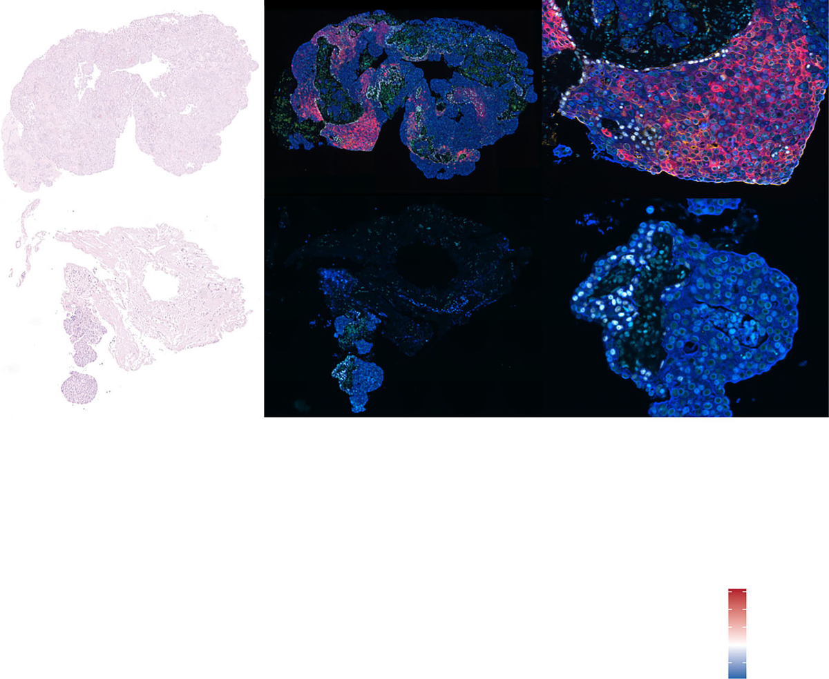
Figure 2. Fluorescent imaging highlights the tumor’s cellular architecture, capturing how specialized cancer cell populations organize within the tissue.
A Blueprint for Functional Precision Oncology in Rare Cancers
The implications of this work extend well beyond SDSC. The study demonstrates a generalizable framework: for rare cancers that lack cell lines, animal models, and clinical trial data, the combination of multimodal molecular profiling with ex vivo tissue slice drug testing can generate biologically informed therapeutic hypotheses from a single surgical specimen. The approach identifies not just whether a drug works, but which specific tumor cell states it eliminates and through which molecular mechanisms — information that is simply unavailable from bulk viability assays or conventional histology.
The validation findings add further weight. The ALDH1A1+ tumor state identified in the index case was present in all 12 SDSC tumors in the independent cohort, with significantly higher expression in recurrent and metastatic samples compared to primary tumors. This suggests that the stem-like ALDH1A1+ compartment may be enriched during disease progression — making its sensitivity to mTOR inhibition all the more clinically relevant.
For the broader precision oncology community, the study offers a practical template: when conventional preclinical models don’t exist, patient-derived tissue slices sectioned with sufficient precision to preserve native architecture can serve as a living laboratory for drug discovery. The Compresstome® platform sits at the center of that workflow, providing the reproducible, artifact-free sections that make downstream single-cell and spatial analyses interpretable.
We are proud to see the Compresstome® contributing to research that could meaningfully change outcomes for patients with SDSC and other rare, poorly understood malignancies. If your lab is working on ex vivo drug response profiling, tissue slice cultures for precision oncology, or spatial biology in human tumor specimens, we’d love to hear about your work. Reach out to our team to discuss how the Compresstome can support your research.