

Formulas and rationale for live sectioning of brain, heart, lung, liver, organoids, and tumors
- There is no universal buffer. Start from six requirements every sectioning buffer shares (isotonic ~300 mOsm, pH 7.3 to 7.4, balanced ions, glucose, oxygenation, ice-cold), then add tissue-specific protectants.
- Brain: carbogen-bubbled aCSF; switch to an NMDG- or sucrose-substituted aCSF for cutting to limit excitotoxic swelling.
- Heart: Tyrode’s solution with a contraction uncoupler (BDM) so the muscle does not tear itself during the cut.
- Lung: inflate with low-melt agarose, then cut in ice-cold HBSS; the agarose gives soft, air-filled tissue enough rigidity to slice.
- Liver: transport in cold UW preservation solution, cut in carbogenated Krebs-Henseleit buffer to protect an ischemia-sensitive organ.
- Organoids and tumors: embed in agarose and cut in ice-cold PBS/medium or HBSS with antibiotics, then recover at an air-liquid interface.
- A vibrating microtome such as the Compresstome sections fresh, unfixed tissue submerged in any of these buffers, with agarose embedding built into the workflow.
Why There Is No Universal Buffer
When you section living tissue, the buffer is not a passive bath. It is life support. For the minutes to hours between removing tissue from its blood supply and getting a slice into culture, the buffer has to do everything the circulation used to do: hold osmotic and ionic balance, supply oxygen and fuel, buffer pH, and limit the metabolic and mechanical damage of cutting. Get it wrong and the readout suffers before the experiment even begins.
Every good sectioning buffer, whatever the tissue, satisfies the same six requirements.
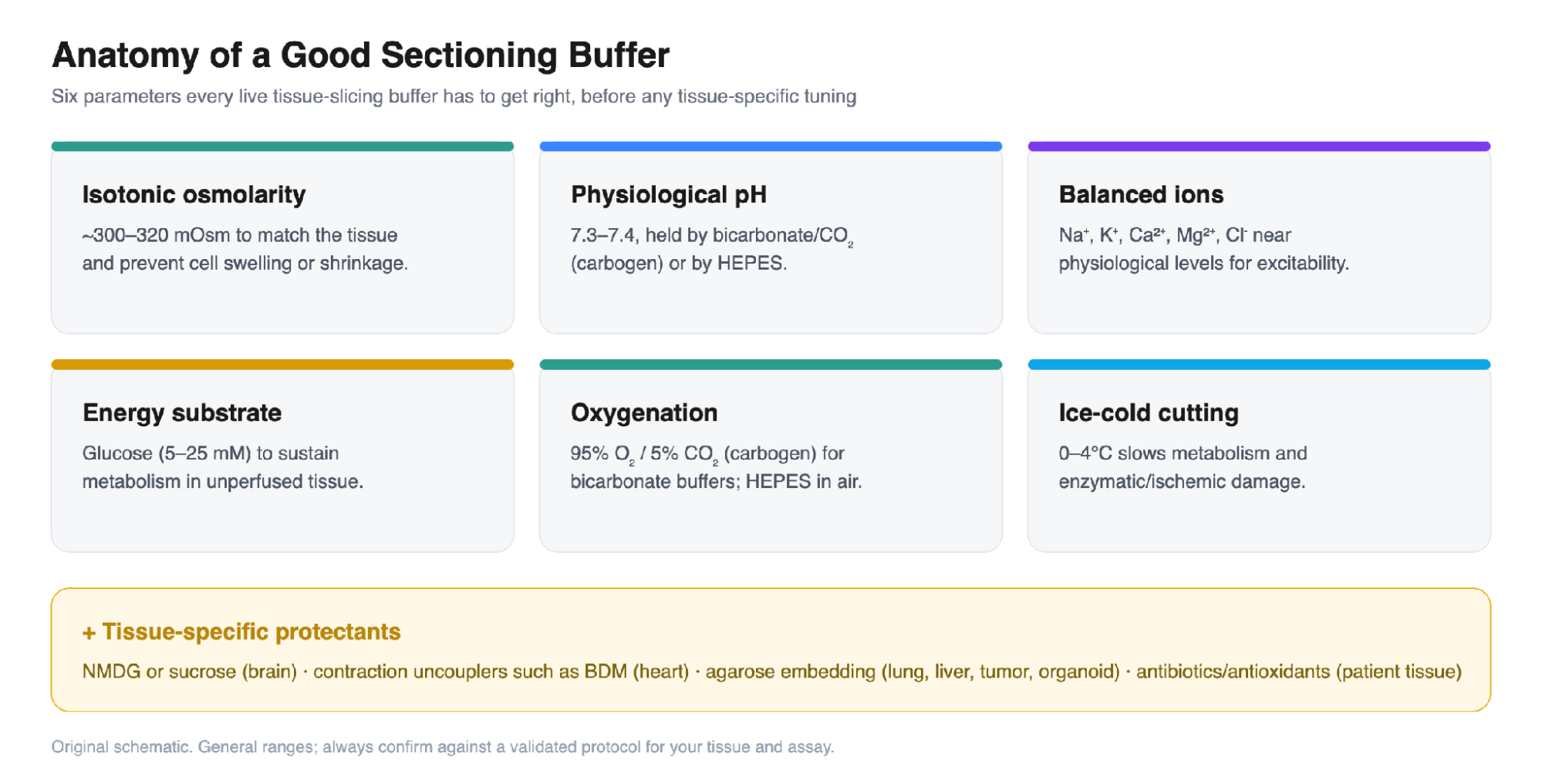
Figure 1. The six parameters shared by any live-tissue sectioning buffer (osmolarity, pH, ionic balance, energy substrate, oxygenation, temperature), plus the tissue-specific protectants layered on top. Original schematic.
Match the Buffer to the Tissue
With those fundamentals in place, the differences between tissues come down to a few decisive additions: how excitable, contractile, soft, or ischemia-sensitive the tissue is. The quick-reference below maps each tissue to its recommended buffer and the single additive that matters most; full compositions and rationale follow.
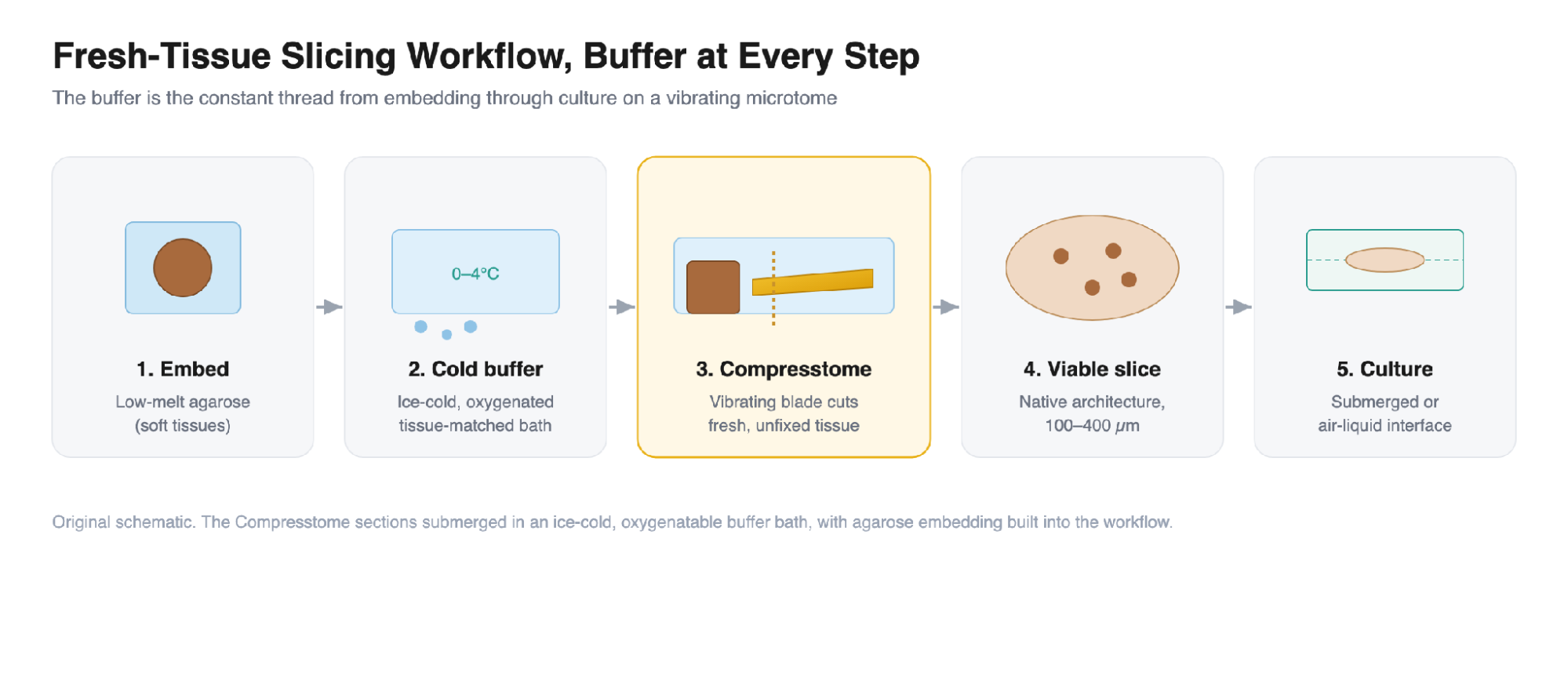
Figure 2. At-a-glance buffer selection for six common sectioning applications, with the additive that most affects slice quality in each. Original schematic; see text for compositions and references.
Brain: artificial cerebrospinal fluid (aCSF)
Acute brain slices are cut and maintained in artificial cerebrospinal fluid, a bicarbonate-buffered salt solution continuously bubbled with carbogen (95% O2 / 5% CO2).
Composition (mM): NaCl 125, KCl 2.5, NaH2PO4 1.25, NaHCO3 25, MgCl2 1, CaCl2 2, glucose 25; carbogen-bubbled; ~300 to 310 mOsm; pH 7.4.
For the cutting step itself, especially in adult or aged brain, a protective NMDG-substituted aCSF markedly improves neuronal survival:
Composition (mM): NMDG 92, KCl 2.5, NaH2PO4 1.25, NaHCO3 30, HEPES 20, glucose 25, sodium ascorbate 5, thiourea 2, sodium pyruvate 3, MgSO4 10, CaCl2 0.5; titrate to pH 7.3 to 7.4 with HCl; carbogen-bubbled.
Rationale: during cutting, neurons are vulnerable to excitotoxicity and osmotic swelling. Replacing Na+ with the impermeant cation NMDG+ limits passive Na+ (and downstream Ca2+) influx; high Mg2+ with low Ca2+ blunts NMDA-receptor excitotoxicity; and ascorbate, pyruvate, and thiourea scavenge reactive oxygen species. A sucrose-substituted aCSF (sucrose replacing NaCl) achieves similar protection (Ting et al., 2018; Ting et al., 2014). See Precisionary’s live brain sectioning application.
Heart: Tyrode’s solution with a contraction uncoupler
Living myocardial slices are prepared in Tyrode’s solution, with tissue handled warm (37 °C, heparinized) and then cut cold (4 °C) while the bath is oxygenated. The decisive addition is an excitation-contraction uncoupler, typically 2,3-butanedione monoxime (BDM).
Composition (mM): NaCl 140, KCl 5.4, CaCl2 1.8, MgCl2 1, glucose 10, HEPES 10; pH 7.4; plus BDM 30 mM during preparation and cutting.
Rationale: myocardium contracts, and an actively twitching block tears under a vibrating blade. BDM reversibly inhibits myosin cross-bridge cycling, mechanically silencing the tissue so it can be cut cleanly and survive. Blebbistatin is a cleaner alternative for the culture phase, since BDM has off-target mitochondrial and phosphatase effects (Watson et al., 2017). See Precisionary’s heart sectioning application.
Lung: HBSS with low-melt agarose inflation
Precision-cut lung slices start by inflating the airways with warm low-melting-point agarose, letting it gel on ice, then coring and cutting in Hank’s Balanced Salt Solution (HBSS).
Composition (mM): Inflation: 1.5 to 2% low-melt agarose in HBSS, warmed to 37 to 42 °C. Cutting: ice-cold HBSS with Ca2+ and Mg2+; ~250 µm slices.
Rationale: air-filled lung is far too compliant to section directly. Instilled agarose is liquid when warm and solid when cold, so it transiently fills the alveoli and airways to provide uniform mechanical support, and it can be melted back out at 37 °C once slices are in culture. HBSS containing Ca2+ and Mg2+ preserves viability and cell adhesion (Li et al., 2022). See Precisionary’s lung sectioning application.
Liver: Krebs-Henseleit buffer with UW solution for transport
Precision-cut liver slices are cut in carbogen-saturated Krebs-Henseleit buffer, with the explant transported and stored cold in University of Wisconsin (UW) preservation solution to minimize ischemia.
Composition (mM): Krebs-Henseleit: NaCl 118, KCl 4.7, CaCl2 2.5, MgSO4 1.2, KH2PO4 1.2, NaHCO3 25, glucose 11; carbogen-saturated; pH 7.4. Cut at 4 °C; ~250 µm slices.
Rationale: the liver is metabolically demanding and highly ischemia-sensitive. UW solution (high K+, lactobionate, raffinose, adenosine, glutathione, allopurinol, hydroxyethyl starch) is a cold-storage preservation fluid designed to suppress cell swelling and oxidative injury during transport; carbogenated Krebs-Henseleit then supplies oxygenation, pH buffering, and substrate through the cut (de Graaf et al., 2010). See Precisionary’s liver sectioning application.
Organoids: ice-cold PBS or medium, agarose, and an air-liquid interface
Mature organoids are embedded in low-melt agarose and cut in ice-cold PBS or culture medium, then recovered at an air-liquid interface on porous membrane inserts.
Composition (mM): Embedding: 2 to 4% low-melt agarose. Cutting: ice-cold PBS or culture medium; 200 to 500 µm (commonly ~250 µm). Recovery: medium plus penicillin-streptomycin-amphotericin, 37 °C.
Rationale: organoids are small and fragile, so an agarose matrix is what makes reproducible cutting possible. Isotonic, buffered PBS or medium preserves viability during the brief cut, and slicing onto an air-liquid interface relieves the hypoxic, necrotic core that otherwise limits large organoids, exposing the interior directly to oxygen and nutrients. Antibiotic and antimycotic coverage is essential for downstream culture (Giandomenico et al., 2019; cortical organoid slice protocol, 2024). See Precisionary’s organoid sectioning application.
Tumors: ice-cold HBSS with antibiotics and agarose embedding
Organotypic tumor slices, including from patient specimens, are transported in ice-cold HBSS with antibiotics, embedded in low-melt agarose, and cut slowly on a vibrating microtome.
Composition (mM): Transport: ice-cold HBSS with Ca2+ and Mg2+, plus antibiotics (± an antioxidant). Embedding: 3 to 5% low-melt agarose. Cutting: ~300 µm at a slow blade advance (0.2 to 0.7 mm/s).
Rationale: patient tumor tissue is heterogeneous, often soft or partly necrotic, and carries contamination risk. Ice-cold HBSS with Ca2+ and Mg2+ plus antibiotics keeps the tissue and its microenvironment (including infiltrating immune cells) viable and sterile; low-melt agarose stabilizes irregular tissue; and a slow blade advance accommodates variable density (Misra et al., 2021). See Precisionary’s tumor sectioning application.
One Instrument, Every Buffer
What these protocols share is more important than what separates them: each cuts fresh, unfixed tissue submerged in a cold, oxygenatable buffer, and most rely on agarose embedding to hold soft tissue steady. That is exactly the workflow a vibrating microtome is built for. The Compresstome sections submerged in an ice-cold buffer bath that you fill with the tissue-appropriate solution, with agarose embedding integrated into the specimen holder, so the same instrument moves cleanly from brain to heart to lung to liver to organoids and tumors. For a broader overview, see precision-cut tissue slices.
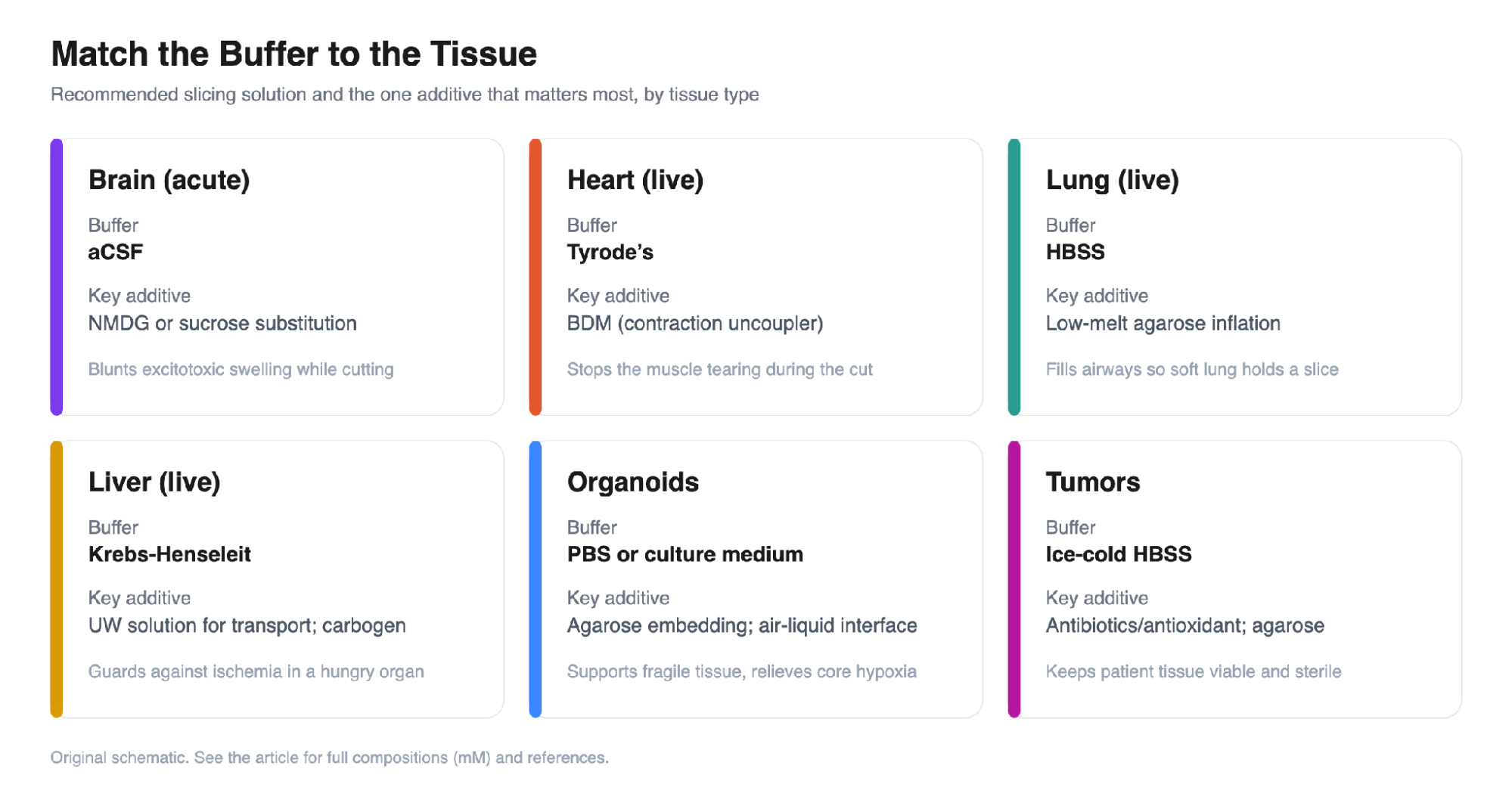
Figure 3. The buffer is the constant thread from agarose embedding, through the ice-cold oxygenated bath and the vibrating-blade cut, to submerged or air-liquid-interface culture. Original schematic.
The takeaway for buffer selection: nail the six fundamentals first, add the one or two protectants your tissue demands, and keep the bath cold and oxygenated through the cut. Do that, and slice quality stops being the variable that limits your experiment.
Not sure which buffer or setup fits your tissue?
Our team helps researchers dial in tissue-specific buffers, embedding, and slicing parameters every day. Tell us your tissue and assay, and we will help you get to viable, reproducible slices faster with the Compresstome vibrating microtome.
→ Browse tissue sectioning help and protocols
→ Explore the Compresstome vibrating microtomes
→ Talk to our team: Contact Us
Email [email protected] Call 617-682-0586