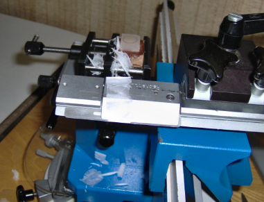
If you work with tissue sectioning regularly, you know how important it is to get clean, consistent slices for your experiments. Whether you’re conducting histology, electrophysiology, or molecular studies, the quality of your tissue sections can make all the difference. But what happens when your sliding microtome starts causing more problems than it solves? Uneven cuts, mechanical failures, and constant maintenance can slow down your workflow and introduce variability into your data. If you’re frustrated with these challenges, it might be time to switch from a sliding microtome to a vibrating microtome, or vibratome. Unlike sliding microtomes, which rely on a stationary blade and a moving tissue block, vibratomes use a gently oscillating blade to minimize compression and preserve tissue integrity. One of the best options available today is the Compresstome vibratome, which provides researchers with precise, reproducible sections while making the slicing process more efficient.
In this article, we’ll explore common problems with sliding microtomes, explain why switching to a vibratome like the Compresstome can improve your research, and provide practical tips for making the transition as smooth as possible.

Common Problems with Sliding Microtomes
Sliding microtomes have been a staple in research and pathology labs for years, but they come with their fair share of limitations. Many researchers find that the manual operation of these microtomes introduces inconsistencies, especially when working with soft or delicate tissues.
One of the biggest challenges is uneven or inconsistent cuts. Because the tissue block moves across the blade manually, even slight variations in pressure can affect the thickness of each section. This inconsistency makes it difficult to obtain reliable results, particularly for imaging and staining applications that require uniform slices.
Another common issue is tissue compression and damage. Sliding microtomes work well for paraffin-embedded samples, but fresh or soft tissues often become distorted under mechanical pressure. This can be a major problem for researchers studying live or lightly fixed samples, where preserving cellular structure is critical.
Maintenance is another concern. Blade sharpening, calibration, and routine adjustments are all necessary to keep a sliding microtome in good working condition. Over time, these maintenance tasks can add up, consuming valuable lab time and resources.
Finally, sliding microtomes are limited in the types of tissue they can handle. They are ideal for harder, paraffin-embedded samples, but struggle with fresh brain, lung, kidney, and other soft tissues. For researchers who need to cut fresh tissue for organotypic culture, electrophysiology, or molecular studies, a sliding microtome may not be the best fit.
Why a Vibratome is the Better Solution
For many researchers, making the switch from a sliding microtome to a vibratome provides an immediate improvement in tissue sectioning. Unlike sliding microtomes, vibratomes use a vibrating blade to gently slice through the tissue, reducing mechanical stress and improving section quality.
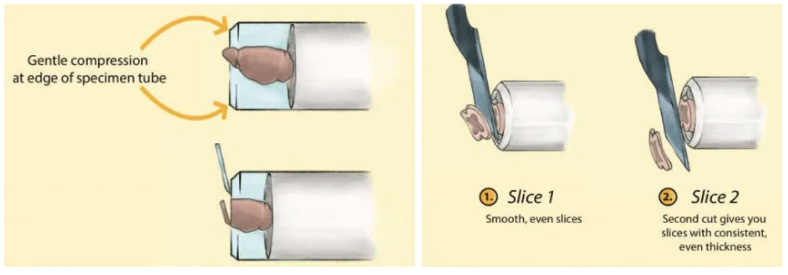
The Compresstome vibratome takes this a step further by incorporating an agarose embedding system, which stabilizes the tissue during slicing. This setup helps researchers achieve thin, even sections with minimal distortion, making it ideal for applications such as electrophysiology, histology, and live tissue imaging.
One of the biggest advantages of using the Compresstome is its ability to preserve tissue integrity. The vibrating blade produces clean cuts without compressing or tearing the sample, ensuring that delicate structures remain intact. This is particularly important for researchers studying live neurons, organoid cultures, or thin tissue sections for imaging.
Another benefit is consistency. The Compresstome allows users to set precise cutting parameters, ensuring that every slice is the same thickness. This level of control leads to more reproducible results, which is essential for high-quality research.

The transition to a vibratome also means less maintenance and user fatigue. Unlike sliding microtomes, which require regular blade sharpening and calibration, the Compresstome is designed for efficiency. Researchers can focus on their experiments rather than troubleshooting equipment.
And finally, the Compresstome is highly versatile. It works well for both fresh and fixed tissues, making it an excellent choice for labs working with a variety of biological samples. Whether you’re studying brain slices, liver sections, or precision-cut lung slices (PCLS), this vibratome offers the flexibility needed for modern research.
Making the Switch: How to Transition from a Sliding Microtome to a Compresstome vibratome
If you’re considering replacing your sliding microtome with a vibratome, the transition is easier than you might think. The key is to adjust your workflow to take full advantage of the vibratome’s capabilities.
First, take time to understand how your specific research applications will benefit from vibratome sectioning. If you’re working with fresh tissue, you’ll quickly see the advantages in cell viability and slice quality. If you’re preparing samples for immunohistochemistry, you’ll notice improved staining uniformity and tissue preservation.
Next, it’s important to train lab members on vibratome operation. While a vibratome is straightforward to use, understanding how to optimize blade oscillation, cutting speed, and agarose embedding techniques will help ensure the best results.
One of the biggest changes for labs switching from a sliding microtome is embedding. While sliding microtomes use paraffin or cryosectioning techniques, vibratomes use low-melt agarose to stabilize tissue samples. Mastering this process is key to getting clean, reproducible sections.
Another consideration is tissue storage. Since vibratomes work well with fresh tissue, it’s important to store samples in ice-cold, oxygenated buffer if they need to be kept viable before slicing. If you’re working with fixed samples, proper fixation and cryoprotection will ensure long-term tissue preservation.
Upgrade Your Tissue Sectioning with the Compresstome
If you’re frustrated with your sliding microtome, switching to a Compresstome vibratome could be the solution you’ve been looking for. Whether you’re working with neuroscience, histology, or organotypic cultures, the Compresstome offers:
- High-quality, consistent tissue sections
- Minimal compression and tissue distortion
- Easy operation with reduced maintenance
- Versatility for fresh and fixed tissue applications
If you’re ready to upgrade your tissue sectioning workflow, our team at Precisionary Instruments is here to help. We specialize in tissue sectioning solutions and can guide you through selecting the right model, optimizing your protocol, and ensuring a smooth transition from your sliding microtome.
Contact us today to learn more about how the Compresstome vibratome can improve your research.