Advancing Neuroscience Through Live Imaging of the Brain’s Extracellular Space
We are honored to spotlight the work of Dr. Virginia Puente, an MSCA and María Zambrano researcher at the University of the Basque Country (UPV/EHU). A pharmacist by training, Dr. Puente leads an ambitious and innovative research program centered on understanding how the extracellular space (ECS) of the brain influences neuronal signaling and synaptic plasticity.
Her work integrates molecular neuroscience, super-resolution microscopy, and optogenetics, with a particular emphasis on visualizing and manipulating neuronal microenvironments in live brain tissue. By developing novel optical tools and implementing refined tissue preparation protocols, Dr. Puente’s research is helping to reveal the dynamic architecture and regulatory function of the ECS—an area of growing importance in the study of neurological disorders and brain health.
Investigating the Structure and Function of the Brain’s Extracellular Space
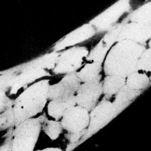
At the core of Dr. Puente’s research is the hypothesis that the ECS is not merely a passive compartment, but rather a structurally dynamic and functionally critical space that modulates the communication between neurons. Her team utilizes Super-resolution Shadow Imaging (SUSHI) and STED microscopy to image the ECS at nanoscale resolution, and combines these techniques with optogenetic constructs that allow targeted activation and manipulation of specific neuronal populations.
Through this work, Dr. Puente is not only mapping ECS architecture in organotypic hippocampal slices, but also developing new tools for probing cell-matrix interactions, diffusion dynamics, and mechanotransduction. These insights have important implications for understanding the pathogenesis of neurological diseases in which ECS remodeling and dysregulation are implicated.

Precision Tissue Preparation Using the Compresstome® VF-510-0Z
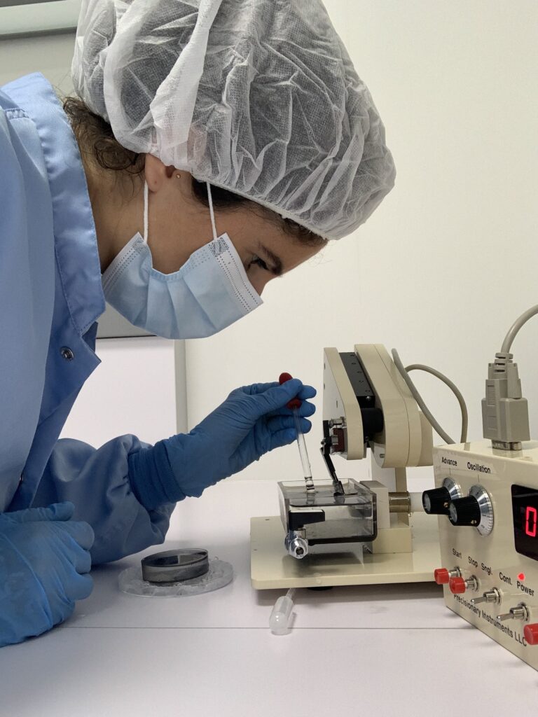
In order to generate high-quality, reproducible slices of mouse hippocampus for her imaging and culture studies, Dr. Puente uses the Compresstome® vibrating microtome from Precisionary Instruments. Her protocol involves embedding dissected hippocampi in 2% agarose within a custom-shaped mold, which is then chilled and mounted in a specimen tube provided with the instrument. The embedded samples are sectioned with high precision, producing viable slices that retain the native architecture of the hippocampus for long-term culture.
“We are really happy using the Compresstome®… It’s made our life much easier and helped us make huge progress compared to our previous setup.”
— Dr. Virginia Puente
Once sectioned, the hippocampal slices are plated using a roller drum culture system, which allows extended in vitro viability—providing a stable platform for long-term imaging and experimental manipulation without the need for repeated animal sacrifice.

Recent Webinar: Tools and Techniques for Live Brain Slice Imaging
We were pleased to host Dr. Puente for a live webinar on April 22, 2025, in which she shared her comprehensive workflow for preparing, culturing, and imaging organotypic hippocampal slices. The webinar covered:
- The step-by-step process of hippocampal tissue dissection and embedding
- Use of the Compresstome® vibratome for precise and consistent sectioning
- Implementation of roller drum culture for extended slice viability
- Super-resolution shadow imaging (SUSHI) and STED microscopy techniques
- Challenges, troubleshooting strategies, and lessons learned
- Optogenetic integration and real-time ECS visualization
Throughout the presentation, Dr. Puente emphasized the importance of protocol standardization and sample integrity, and highlighted the role that reliable tissue sectioning plays in the success of complex imaging studies.
View the Full Recording
To gain further insight into Dr. Puente’s work and techniques, we invite you to view the full webinar recording:
Watch the Full Webinar Recording
If you are interested in implementing similar experimental approaches in your own research, or would like more information about the Compresstome® vibratome, please don’t hesitate to contact our team at [email protected].