Lab Spotlight: The TAB-Lab at The Hong Kong University of Science and Technology
- Original post date:
Share on social
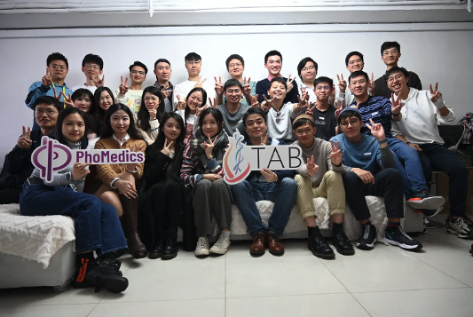
In this Lab Spotlight, we’re thrilled to feature the groundbreaking work of the Translational and Advanced Bioimaging Laboratory (TAB-Lab), led by Dr. Terence Wong at The Hong Kong University of Science and Technology (Figure 1). This innovative team is redefining the field of bioimaging with cutting-edge tools and techniques that bridge the gap between basic research and clinical applications.

Pioneering Advances in Bioimaging
At the Translational and Advanced Bioimaging Laboratory (TAB-Lab) at The Hong Kong University of Science and Technology, Principal Investigator Dr. Terence Wong and his team are pushing the boundaries of biological imaging technology. Their research focuses on developing state-of-the-art 2D and 3D imaging systems, synthesizing biomarkers, and designing deep learning algorithms that enhance both fundamental life science research and biomedical applications.
One of their most impactful innovations is CHAMP, a high-speed computational microscope that, when combined with a weakly-supervised deep learning algorithm, enables virtual histological staining of unprocessed tissue in just three minutes. This technology offers an exciting step forward in real-time cancer diagnostics, reducing the need for secondary surgeries and improving patient outcomes.
Beyond cancer diagnostics, the TAB-Lab has also developed whole-organ fluorescence imaging systems using block-face imaging with serial sectioning. Their TRUST, HistoTRUST, and HiLoTRUST technologies allow for high-content, subcellular-resolution imaging of entire mouse organs or embryos, making significant contributions to developmental biology and 3D histology (Figure 2).

The Role of the Compresstome Vibratome in the TAB-Lab

A key tool in the lab’s imaging pipeline is the Compresstome vibratome, which plays a crucial role in their serial sectioning techniques. The precision, distortion-free slicing provided by the vibratome is essential for whole-organ imaging methods such as TRUST, HistoTRUST, and HiLoTRUST.
By using the Compresstome, the TAB-Lab can generate thin, uniform tissue sections, ensuring that imaging data remains accurate and high-resolution. This capability is particularly vital when studying entire biological specimens, as it allows researchers to create detailed, high-quality 3D reconstructions of organs and tissues (Figure 3).
Shaping the Future of Biomedical Imaging
Through groundbreaking work in computational microscopy, deep learning applications, and whole-organ imaging, the TAB-Lab is setting new standards for non-invasive, high-resolution tissue imaging. Their use of innovative tools like the Compresstome vibratome helps bridge the gap between basic research and clinical applications, paving the way for more efficient diagnostics and deeper biological insights.
The impact of Dr. Wong’s team extends beyond research—they are driving advancements that will transform the way we visualize, analyze, and understand biological tissues. As their work progresses, the future of bioimaging and tissue diagnostics looks brighter than ever.
Advancing Bioimaging with Precision Tissue Sectioning
The innovative research happening at the TAB-Lab highlights the importance of precise tissue sectioning for whole-organ imaging and biomedical applications. The Compresstome vibratome plays a crucial role in their workflows, enabling distortion-free sectioning for high-resolution imaging techniques like TRUST and HiLoTRUST.
If your lab is working on advanced bioimaging, serial sectioning, or tissue preparation for 3D histology, explore how the Compresstome vibratome can enhance your research. Learn more about our vibratomes here, or reach out to our team for expert guidance on selecting the right model for your needs!
Share on social
