Advancing skin research depends on high-quality tissue samples, and a recent study (Ziolkowski et al. from Science Advances) is a great example of how innovative sectioning techniques can make a big difference. This team of researchers set out to better understand touch detection in skin tissue, and to do that, they needed clear, precise sections that preserved tissue integrity. Their solution? The Compresstome vibratome, which helped them achieve smooth, reproducible slices without the usual artifacts that come with traditional methods.
Getting a Clearer Picture of Skin Tissue
Tissue sectioning is a critical step in many types of research, from studying disease pathology to testing new therapies. But it’s not always easy to get high-quality sections, especially with fragile samples like skin. Conventional methods—like paraffin embedding or cryosectioning—often introduce compression artifacts, tearing, or freezing damage. For this study, the researchers wanted to avoid these issues, so they used the Compresstome vibratome to ensure uniform, high-quality slices.
With these precise sections, they were able to perform histological staining and imaging, giving them a clear and detailed look at how skin cells were organized and how specific biomarkers were distributed. Their findings have important implications for wound healing, dermatology, and regenerative medicine, offering insights that could help develop better treatments for skin disorders.
Key Findings and Why They Matter
One of the major takeaways from this study was that high-quality tissue sections lead to better data. The researchers found that immunofluorescent staining showed stronger and more defined biomarker signals, making it easier to analyze protein expression and cellular structures. Because the sections were consistent across different samples, their data was also more reliable and reproducible, a crucial factor in scientific research.
These results don’t just benefit this one study—they have much broader applications. Consistently high-quality sections are essential for drug testing, regenerative medicine, and dermatopathology. By refining tissue sectioning techniques, researchers can gain a more accurate understanding of disease progression, treatment responses, and new therapeutic targets.
How Precision Sectioning Made a Difference
One of the highlights of this study was the use of precision sectioning tools to improve the quality of tissue slices. With the Compresstome vibratome, the researchers were able to control section thickness while minimizing mechanical distortions. This allowed them to preserve the three-dimensional structure of skin tissue, which is often lost in traditional sectioning methods.

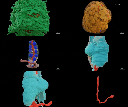
Figure 1. 3D architecture of the Pacinian corpuscle obtained using eFIB-SEM.
By maintaining tissue integrity, the team could perform more accurate histology, immunohistochemistry, and imaging-based studies. Their work highlights how precision sectioning techniques can enhance research outcomes and lead to more meaningful discoveries in dermatology and beyond.
What This Means for Future Research
This study is just one example of how improving tissue sectioning can have a major impact on scientific discoveries. Moving forward, researchers studying wound healing, skin regeneration, and inflammatory conditions can apply these techniques to gain clearer, more reliable data. Additionally, high-quality sections play an important role in tissue engineering, helping scientists develop better skin grafts and test potential new treatments.
As research in dermatology continues to evolve, the demand for better, more reliable tissue sectioning will only grow. This study underscores the importance of precision tools like the Compresstome vibratome in advancing our understanding of skin biology and improving medical research. It’s exciting to see how innovations in tissue preparation can lead to real-world breakthroughs in skin health and therapy development.
Curious to see how precision tissue sectioning can enhance your research? Learn more about the Compresstome vibratome and how it’s helping scientists achieve high-quality, reproducible results. Contact us today to explore how our technology can support your work.