Electrophysiology is the study of the electrical properties of biological cells and tissues, and how these properties generate and propagate electrical signals in the body. This field is used to understand the electrical activity of cells in the heart, brain, and other organs, and to diagnose and treat various health conditions, such as arrhythmias and neurological disorders. Techniques used in electrophysiology include electrocardiograms (ECGs), electroencephalography (EEG), and invasive procedures such as cardiac catheterization.
How does electrophysiology help us study the brain?
Electrophysiology helps us study the brain by recording the electrical activity of neurons.
This is done using techniques such as:
Electroencephalography (EEG): A non-invasive method that records brain activity by placing electrodes on the scalp.
Electrocorticography (ECoG): A less invasive method that records brain activity by placing electrodes directly on the surface of the brain.
Intracranial EEG (iEEG): An invasive method that involves placing electrodes directly into the brain tissue
These techniques allow us to measure the electrical signals produced by neurons, giving us information about the functioning of different brain regions and the timing and coordination of neural activity (Figure 1). This information helps us understand the neural basis of various brain processes such as perception, attention, and memory. It also provides insight into conditions such as epilepsy, Parkinson’s disease, and depression.
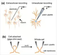
Figure 1. Types of electrical recordings done in the brain.
How does electrophysiology help us study neurons?
Electrophysiology is used to study neurons by measuring the electrical signals generated by their activity. This is done using techniques such as:
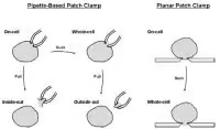
- Patch-clamp recording: A highly invasive method that involves attaching a microscopic glass pipette to a single neuron to measure its electrical signals. There are multiple types of patch-clamp electrophysiology that one can do (Figure 2).
- Multi-electrode recording: A method that involves recording the electrical signals from multiple neurons simultaneously using electrodes placed directly onto brain tissue.
- Optical imaging: A method that uses indicators sensitive to electrical activity and light to visualize the electrical signals generated by neurons.
These techniques allow us to measure the electrical activity of neurons, giving us information about their excitability, firing patterns, and synaptic connections. This information helps us understand the electrical signaling mechanisms underlying neural communication and the underlying basis of various brain processes such as perception, attention, and memory.
How does one do patch-clamp electrophysiology?
Patch-clamp electrophysiology is a technique used to study the electrical activity of individual cells by making a very small and precise seal between the cell and a recording electrode. Here’s a general outline of the steps involved:
- Cell preparation: Choose cells for recording and prepare them for recording (such as isolation of neurons from tissue or culturing cells).
- Cell identification: Observe cells under a microscope and choose the target cell for recording.
- Seal formation: Bring the tip of a glass micropipette near the cell membrane and apply suction to form a tight seal between the pipette and the cell membrane.
- Patch formation: Gently break the cell membrane to create an opening into the cell, forming a “patch.”
- Record signals: Connect the recording electrode to a high-impedance amplifier and record the electrical signals generated by the cell.
- Data analysis: Analyze the recorded data to study the cell’s electrical activity.
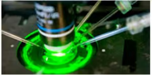
It’s important to note that patch-clamp is a complex technique that requires specialized equipment and training. It should be performed by experienced technicians in a well-equipped laboratory (Figure 3).
Why is patch-clamp electrophysiology important?
Patch-clamp electrophysiology is important for several reasons, including:
- Study of ion channels: The technique allows for the study of ion channels, which play crucial roles in cell signaling, electrical signaling in the nervous system, and muscle contraction.
- Single-cell analysis: The technique enables the study of single cells, which can provide more detailed and precise information compared to conventional whole-cell recordings.
- Determination of channel properties: The technique can be used to determine important properties of ion channels such as their conductance, kinetics, and sensitivity to drugs or other modulators.
- Identification of disease-causing mutations: The technique can be used to identify mutations that cause ion channel dysfunction, which can lead to various diseases such as epilepsy, cardiac arrhythmias, and some types of inherited deafness.
- Development of new drugs: The technique can be used to test the efficacy and mechanism of action of new drugs that target ion channels, which is important for the development of new treatments for various diseases.
Overall, patch-clamp electrophysiology provides a valuable tool for understanding the function of ion channels and their roles in normal and abnormal cellular processes.
How is a vibratome used in preparing tissue slices for electrophysiology?
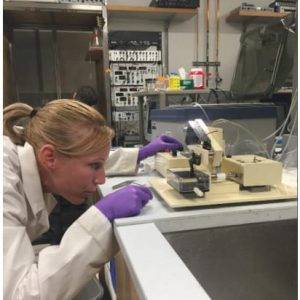
A vibratome is a necessary component for patch-clamp electrophysiology, because it can be used to prepare thin slices of tissues, such as brain slices, for recording. The use of a vibratome (Figure 4) can be important in certain types of patch-clamp experiments because it allows for the study of cells in their native tissue environment, rather than in dissociated or cultured cells.
Benefits of using a vibratome to prepare tissue slices for electrophysiology include:
- Preparation of thin slices: The vibratome uses a rapidly vibrating blade to cut thin slices of tissue, which can then be used for recording.
- Maintenance of cellular and synaptic connections: Thin slices maintain the cellular and synaptic connections between cells, which can provide a more physiologically relevant preparation for recording.
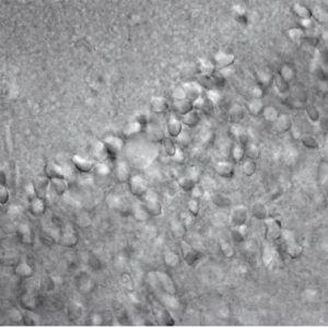
- Study of synaptic transmission: The use of thin slices can allow for the study of synaptic transmission, which is the transfer of electrical or chemical signals from one neuron to another (Figure 5).
Overall, patch-clamp electrophysiology provides a valuable tool for understanding the function of ion channels and their roles in normal and abnormal cellular processes.
Overall, a vibratome can provide advantages for certain types of experiments by allowing for the study of cells in their native tissue environment.
