

Exciting Research Update from Stanford University’s Madison Lab!As the global community of Compresstome® vibratome users continues to expand, we are thrilled to spotlight the groundbreaking research of Professor Daniel Madison and his team at Stanford University.
The Compresstome® Vibratome in the Madison Lab:
The Madison Lab specializes in exploring the fundamental properties, plasticity, and modulation of central nervous system synapses. Their work includes studying the detailed structure and protein content of synapses in different plastic states, as well as investigating the pathophysiology of Alzheimer’s disease in relation to endocannabinoids.
Utilizing a wide range of electrophysiological techniques, including extracellular field potential recording, intracellular recording, and whole-cell/single-channel recording in hippocampal slices and cultured neurons, the lab delves into the intricate mechanisms underlying synaptic function and plasticity.
Their recent projects encompass a diverse range of topics, such as the role of the amyloid peptide A-beta in modulating synaptic inhibition, the involvement of the Fragile X mental retardation protein in neural circuit formation, and an array tomographic study on synaptic plasticity’s influence on neural microcircuits.
🌟 Notably, the Madison Lab has incorporated the Compresstome® VF-300-0Z vibratome into their research toolkit. “We have only been using the new Compresstome for a few weeks, but have noted an immediate improvement in the quality of our brain slices,” reports Dr. Madison. “In particular, the slices have ‘cleaner’ surfaces and the neurons seem healthier.”
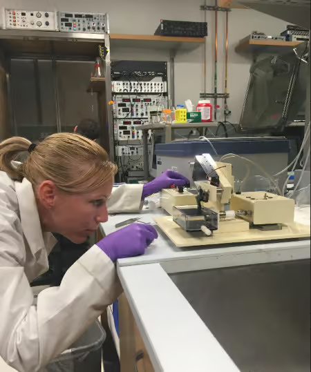
Here is lab member Dr. Marianna Kiraly preparing mouse hippocampal slices on our Compresstome vibratome.
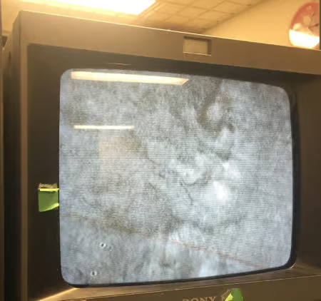
After making acute mouse brain sections, here is what the neurons look like: A photograph of a TV screen on Dr. Kiraly’s electrophysiological rig of the surface of the Compresstome®-prepared slices. The lab uses these TV images to target neurons for the electrophysiological electrodes.
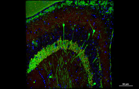
Last but not least, here is a beautiful volume reconstruction image of a mouse hippocampal slice, taken from a YFP (yellow fluorescent protein) H-line mouse (green cells), also immunostained for DAPI (blue) and synaptophysin (red). Slice and photo credit: Dr. Ricardo Valenzuela, a former lab member. Our Compresstome vibratome is capable of sectioning brain slices for both electrophysiology and immunohistochemistry — all on one machine.
Find out more about the Madison Lab at Stanford!
Questions about generating healthy acute mouse brain slices for electrophysiology or about patch-clamping experiments? Reach out to our scientific experts at Precisionary Instruments directly.